El-Sayed H Ibrahim, Lubna Chaudhary, Yee-Chung Cheng, Antonio Sosa, Dayeong An, John Charlson
{"title":"Cardiac MRI for differentiating chemotherapy-induced cardiotoxicity in sarcoma and breast cancer.","authors":"El-Sayed H Ibrahim, Lubna Chaudhary, Yee-Chung Cheng, Antonio Sosa, Dayeong An, John Charlson","doi":"10.2478/raon-2025-0012","DOIUrl":null,"url":null,"abstract":"<p><strong>Background: </strong>Over the past few decades, many studies have focused on anthracyclines effect on the heart (cardiotoxicity), but only a few have focused on sarcoma. In this study, we harness the capabilities of advanced cardiac magnetic resonance imaging (MRI) for characterizing anthracyclines-induced cardiotoxicity in sarcoma and compare the results to those from breast cancer patients.</p><p><strong>Patients and methods: </strong>The patients receive an MRI exam at three timepoints: baseline (pre-treatment), posttreatment, and at 6-months follow-up.</p><p><strong>Results: </strong>The results demonstrated a differential response in sarcoma, characterized by increasing left-ventricular (LV) mass and decreasing right ventricular ejection fraction (RVEF). In all patients, left ventricular ejection fraction (LVEF) remained > 50% at all timepoints. Myocardial strain was always lower than the normal threshold values and showed small changes between different timepoints. Myocardial T2 and extracellular volume (ECV) showed increasing and decreasing patterns, respectively, in sarcoma, which were the opposite patterns of those in breast cancer. While myocardium T1 showed increasing values in breast cancer, T1 in sarcoma increased post-treatment and then decreased at the 6-months follow-up. The results showed inverse correlation between dose and different strain components in sarcoma, which was not the case in breast cancer. Certain myocardial segments showed high correlation coefficients with dose, which may reflect their increased sensitivity to cardiotoxicity.</p><p><strong>Conclusions: </strong>Cardiac MRI proved to be a valuable technique for determining anthracycline-induced changes in cardiac function and myocardial tissue composition in sarcoma and differentiating it against breast cancer. It also provides a comprehensive assessment of heart health at baseline, which is important for risk stratification.</p>","PeriodicalId":21034,"journal":{"name":"Radiology and Oncology","volume":"59 1","pages":"79-90"},"PeriodicalIF":2.2000,"publicationDate":"2025-02-27","publicationTypes":"Journal Article","fieldsOfStudy":null,"isOpenAccess":false,"openAccessPdf":"https://www.ncbi.nlm.nih.gov/pmc/articles/PMC11867570/pdf/","citationCount":"0","resultStr":null,"platform":"Semanticscholar","paperid":null,"PeriodicalName":"Radiology and Oncology","FirstCategoryId":"3","ListUrlMain":"https://doi.org/10.2478/raon-2025-0012","RegionNum":4,"RegionCategory":"医学","ArticlePicture":[],"TitleCN":null,"AbstractTextCN":null,"PMCID":null,"EPubDate":"2025/3/1 0:00:00","PubModel":"eCollection","JCR":"Q3","JCRName":"ONCOLOGY","Score":null,"Total":0}
引用次数: 0
Abstract
Background: Over the past few decades, many studies have focused on anthracyclines effect on the heart (cardiotoxicity), but only a few have focused on sarcoma. In this study, we harness the capabilities of advanced cardiac magnetic resonance imaging (MRI) for characterizing anthracyclines-induced cardiotoxicity in sarcoma and compare the results to those from breast cancer patients.
Patients and methods: The patients receive an MRI exam at three timepoints: baseline (pre-treatment), posttreatment, and at 6-months follow-up.
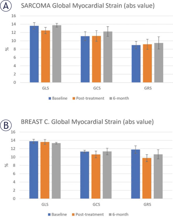
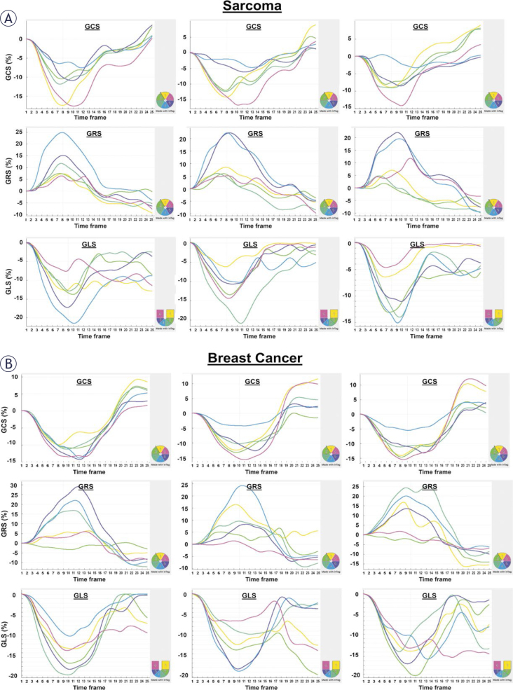
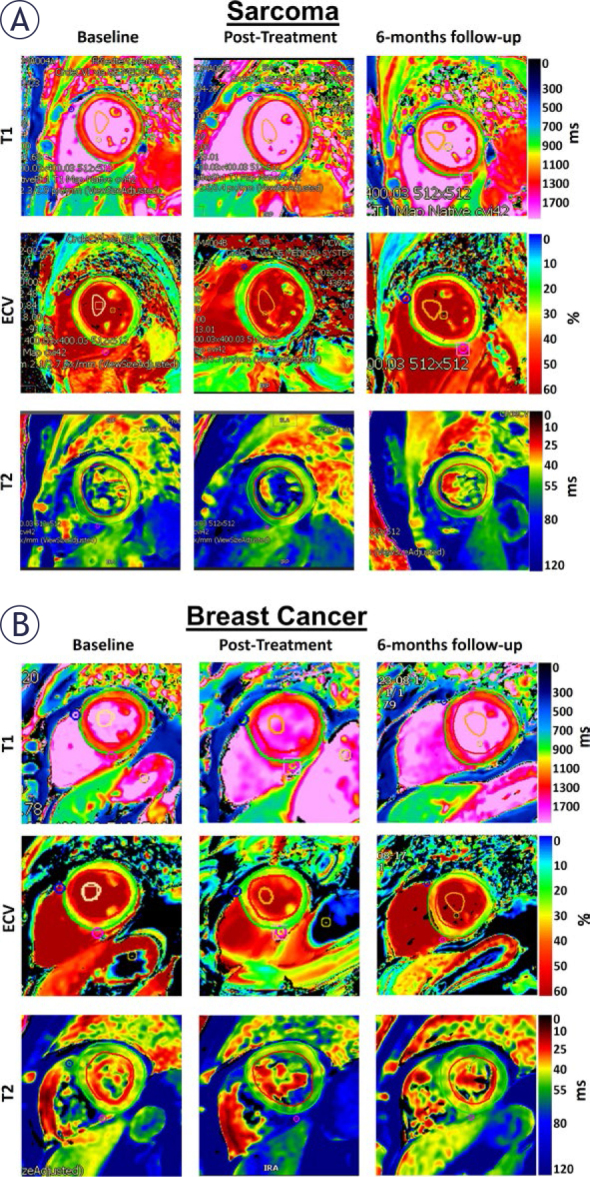
Results: The results demonstrated a differential response in sarcoma, characterized by increasing left-ventricular (LV) mass and decreasing right ventricular ejection fraction (RVEF). In all patients, left ventricular ejection fraction (LVEF) remained > 50% at all timepoints. Myocardial strain was always lower than the normal threshold values and showed small changes between different timepoints. Myocardial T2 and extracellular volume (ECV) showed increasing and decreasing patterns, respectively, in sarcoma, which were the opposite patterns of those in breast cancer. While myocardium T1 showed increasing values in breast cancer, T1 in sarcoma increased post-treatment and then decreased at the 6-months follow-up. The results showed inverse correlation between dose and different strain components in sarcoma, which was not the case in breast cancer. Certain myocardial segments showed high correlation coefficients with dose, which may reflect their increased sensitivity to cardiotoxicity.
Conclusions: Cardiac MRI proved to be a valuable technique for determining anthracycline-induced changes in cardiac function and myocardial tissue composition in sarcoma and differentiating it against breast cancer. It also provides a comprehensive assessment of heart health at baseline, which is important for risk stratification.
期刊介绍:
Radiology and Oncology is a multidisciplinary journal devoted to the publishing original and high quality scientific papers and review articles, pertinent to diagnostic and interventional radiology, computerized tomography, magnetic resonance, ultrasound, nuclear medicine, radiotherapy, clinical and experimental oncology, radiobiology, medical physics and radiation protection. Therefore, the scope of the journal is to cover beside radiology the diagnostic and therapeutic aspects in oncology, which distinguishes it from other journals in the field.


 分享
分享
 求助内容:
求助内容: 应助结果提醒方式:
 扫码关注我们
