Nezka Hribernik, Katja Strasek, Andrej Studen, Katarina Zevnik, Katja Skalic, Robert Jeraj, Martina Rebersek
{"title":"Early-time-point <sup>18</sup>F-FDG-PET/CT and other prognostic biomarkers of survival in metastatic melanoma patients receiving immunotherapy.","authors":"Nezka Hribernik, Katja Strasek, Andrej Studen, Katarina Zevnik, Katja Skalic, Robert Jeraj, Martina Rebersek","doi":"10.2478/raon-2025-0014","DOIUrl":null,"url":null,"abstract":"<p><strong>Background: </strong>A considerable proportion of metastatic melanoma (mM) patients do not respond to immune checkpoint inhibitors (ICIs). There is a great need to develop noninvasive biomarkers to detect patients, who do not respond to ICIs early during the course of treatment. The aim of this study was to evaluate the role of early [<sup>18</sup>F]2fluoro-2-deoxy-D-glucose PET/CT (<sup>18</sup>F-FDG PET/CT) at week four (W4) and other possible prognostic biomarkers of survival in mM patients receiving ICIs.</p><p><strong>Patients and methods: </strong>. In this prospective noninterventional clinical study, mM patients receiving ICIs regularly underwent <sup>18</sup>F-FDG PET/CT: at baseline, at W4 after ICI initiation, at week sixteen and every 16 weeks thereafter. The tumor response to ICIs at W4 was assessed via modified European Organisation for Research and Treatment of Cancer (EORTC) criteria. Patients with progressive metabolic disease (PMD) were classified into the no clinical benefit group (no-CB), and those with other response types were classified into the clinical benefit group (CB). The primary end point was survival analysis on the basis of the W4 <sup>18</sup>F-FDG PET/CT response. The secondary endpoints were survival analysis on the basis of LDH, the number of metastatic localizations, and immune-related adverse events (irAEs). Kaplan-Meier analysis and univariate Cox regression analysis were used to assess the impact on survival.</p><p><strong>Results: </strong>Overall, 71 patients were included. The median follow-up was 37.1 months (952% CI = 30.1-38.0). Three (4%) patients had only baseline scans due to rapid disease progression and death prior to W4 <sup>18</sup>F-FDG-PET/CT. Fifty-one (72%) patients were classified into the CB group, and 17 (24%) were classified into the no-CB group. There was a statistically significant difference in median overall survival (OS) between the CB group (median OS not reached [NR]; 95% CI = 17.8 months - NR) and the no-CB group (median OS 6.2 months; 95% CI = 4.6 months - NR; p = 0.003). Univariate Cox analysis showed HR of 0.4 (95% CI = 0.18 - 0.72; p = 0.004). median OS was also significantly longer in the group with normal serum LDH levels and the group with irAEs and cutaneous irAEs.</p><p><strong>Conclusions: </strong>Evaluation of mM patients with early <sup>18</sup>F-FDG-PET/CT at W4, who were treated with ICIs, could serve as prognostic imaging biomarkers. Other recognized prognostic biomarkers were the serum LDH level and occurrence of cutaneous irAEs.</p>","PeriodicalId":21034,"journal":{"name":"Radiology and Oncology","volume":"59 1","pages":"43-53"},"PeriodicalIF":2.2000,"publicationDate":"2025-02-27","publicationTypes":"Journal Article","fieldsOfStudy":null,"isOpenAccess":false,"openAccessPdf":"https://www.ncbi.nlm.nih.gov/pmc/articles/PMC11867565/pdf/","citationCount":"0","resultStr":null,"platform":"Semanticscholar","paperid":null,"PeriodicalName":"Radiology and Oncology","FirstCategoryId":"3","ListUrlMain":"https://doi.org/10.2478/raon-2025-0014","RegionNum":4,"RegionCategory":"医学","ArticlePicture":[],"TitleCN":null,"AbstractTextCN":null,"PMCID":null,"EPubDate":"2025/3/1 0:00:00","PubModel":"eCollection","JCR":"Q3","JCRName":"ONCOLOGY","Score":null,"Total":0}
引用次数: 0
Abstract
Background: A considerable proportion of metastatic melanoma (mM) patients do not respond to immune checkpoint inhibitors (ICIs). There is a great need to develop noninvasive biomarkers to detect patients, who do not respond to ICIs early during the course of treatment. The aim of this study was to evaluate the role of early [18F]2fluoro-2-deoxy-D-glucose PET/CT (18F-FDG PET/CT) at week four (W4) and other possible prognostic biomarkers of survival in mM patients receiving ICIs.
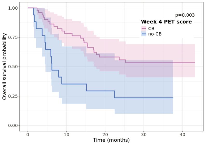
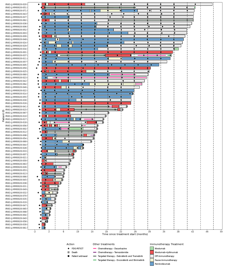
Patients and methods: . In this prospective noninterventional clinical study, mM patients receiving ICIs regularly underwent 18F-FDG PET/CT: at baseline, at W4 after ICI initiation, at week sixteen and every 16 weeks thereafter. The tumor response to ICIs at W4 was assessed via modified European Organisation for Research and Treatment of Cancer (EORTC) criteria. Patients with progressive metabolic disease (PMD) were classified into the no clinical benefit group (no-CB), and those with other response types were classified into the clinical benefit group (CB). The primary end point was survival analysis on the basis of the W4 18F-FDG PET/CT response. The secondary endpoints were survival analysis on the basis of LDH, the number of metastatic localizations, and immune-related adverse events (irAEs). Kaplan-Meier analysis and univariate Cox regression analysis were used to assess the impact on survival.
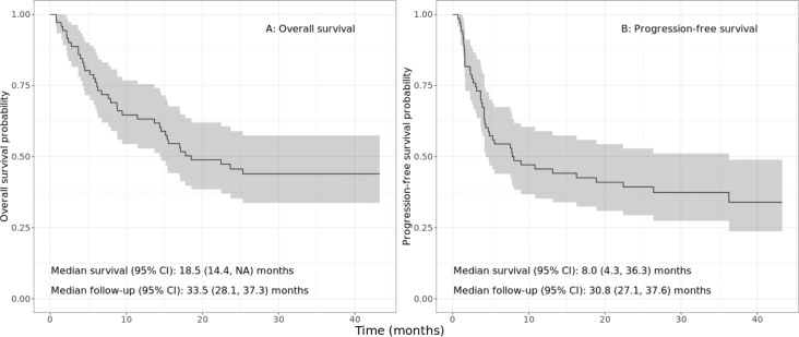
Results: Overall, 71 patients were included. The median follow-up was 37.1 months (952% CI = 30.1-38.0). Three (4%) patients had only baseline scans due to rapid disease progression and death prior to W4 18F-FDG-PET/CT. Fifty-one (72%) patients were classified into the CB group, and 17 (24%) were classified into the no-CB group. There was a statistically significant difference in median overall survival (OS) between the CB group (median OS not reached [NR]; 95% CI = 17.8 months - NR) and the no-CB group (median OS 6.2 months; 95% CI = 4.6 months - NR; p = 0.003). Univariate Cox analysis showed HR of 0.4 (95% CI = 0.18 - 0.72; p = 0.004). median OS was also significantly longer in the group with normal serum LDH levels and the group with irAEs and cutaneous irAEs.
Conclusions: Evaluation of mM patients with early 18F-FDG-PET/CT at W4, who were treated with ICIs, could serve as prognostic imaging biomarkers. Other recognized prognostic biomarkers were the serum LDH level and occurrence of cutaneous irAEs.
期刊介绍:
Radiology and Oncology is a multidisciplinary journal devoted to the publishing original and high quality scientific papers and review articles, pertinent to diagnostic and interventional radiology, computerized tomography, magnetic resonance, ultrasound, nuclear medicine, radiotherapy, clinical and experimental oncology, radiobiology, medical physics and radiation protection. Therefore, the scope of the journal is to cover beside radiology the diagnostic and therapeutic aspects in oncology, which distinguishes it from other journals in the field.


 分享
分享
 求助内容:
求助内容: 应助结果提醒方式:
 扫码关注我们
