Fiona Dierksen, Johanna S Geibel, Janika Albrecht, Sabine Hofer, Peter Dechent, Amelie C Hesse, Jens Frahm, Mathias Bähr, Jan C Koch, Jan Liman, Ilko L Maier
{"title":"T1-relaxation times along the corticospinal tract as a diagnostic marker in patients with amyotrophic lateral sclerosis.","authors":"Fiona Dierksen, Johanna S Geibel, Janika Albrecht, Sabine Hofer, Peter Dechent, Amelie C Hesse, Jens Frahm, Mathias Bähr, Jan C Koch, Jan Liman, Ilko L Maier","doi":"10.3389/fnimg.2025.1549727","DOIUrl":null,"url":null,"abstract":"<p><strong>Background and purpose: </strong>In the differential diagnostic workup of amyotrophic lateral sclerosis (ALS), magnetic resonance imaging (MRI) is primarily used to rule out significant differential diagnoses. So far, whole-brain T1-mapping has not been assessed as a diagnostic tool in this patient population.</p><p><strong>Methods: </strong>We investigated the diagnostic potential of a novel T1-mapping method based on real-time MRI with 0.5 mm in-plane resolution and 4s acquisition time per slice. The study included patients aged 18 to 90 years who met the revised El Escorial criteria for at least possible ALS. T1-relaxation times were measured along the corticospinal tract in predefined regions of interest.</p><p><strong>Results: </strong>Twenty-nine ALS-patients and 43 control group patients (CG) were included in the study. Median ALS Functional Rating Scale revised (ALSFRS-R) was 37 (IQR, 35-44) points and the mean duration from symptom onset to MRI was 21 ± 17 (SD) months. ALS patients showed significantly higher T1-relaxation times in all ROIs compared to CG with mean differences in the hand knob of 50 ms (<i>p</i> < 0.001), corona radiata 24 ms (<i>p</i> = 0.034), internal capsule 27 ms (<i>p</i> = 0.002) and midbrain peduncles 48 ms (<i>p</i> < 0.001). There was a consistent negative correlation between the ALSFRS-R and T1-relaxation times in all ROIs.</p><p><strong>Conclusions: </strong>T1-relaxation times along the corticospinal tract are significantly elevated in ALS patients compared to CG and associated with lower ALSFRS-R. These results imply the analysis of T1-relaxation times as a promising diagnostic tool that can distinguish ALS patients from the control group. Ongoing longitudinal studies may provide deeper insights into disease progression and the effects of therapeutic interventions.</p>","PeriodicalId":73094,"journal":{"name":"Frontiers in neuroimaging","volume":"4 ","pages":"1549727"},"PeriodicalIF":0.0000,"publicationDate":"2025-02-13","publicationTypes":"Journal Article","fieldsOfStudy":null,"isOpenAccess":false,"openAccessPdf":"https://www.ncbi.nlm.nih.gov/pmc/articles/PMC11865248/pdf/","citationCount":"0","resultStr":null,"platform":"Semanticscholar","paperid":null,"PeriodicalName":"Frontiers in neuroimaging","FirstCategoryId":"1085","ListUrlMain":"https://doi.org/10.3389/fnimg.2025.1549727","RegionNum":0,"RegionCategory":null,"ArticlePicture":[],"TitleCN":null,"AbstractTextCN":null,"PMCID":null,"EPubDate":"2025/1/1 0:00:00","PubModel":"eCollection","JCR":"","JCRName":"","Score":null,"Total":0}
引用次数: 0
Abstract
Background and purpose: In the differential diagnostic workup of amyotrophic lateral sclerosis (ALS), magnetic resonance imaging (MRI) is primarily used to rule out significant differential diagnoses. So far, whole-brain T1-mapping has not been assessed as a diagnostic tool in this patient population.
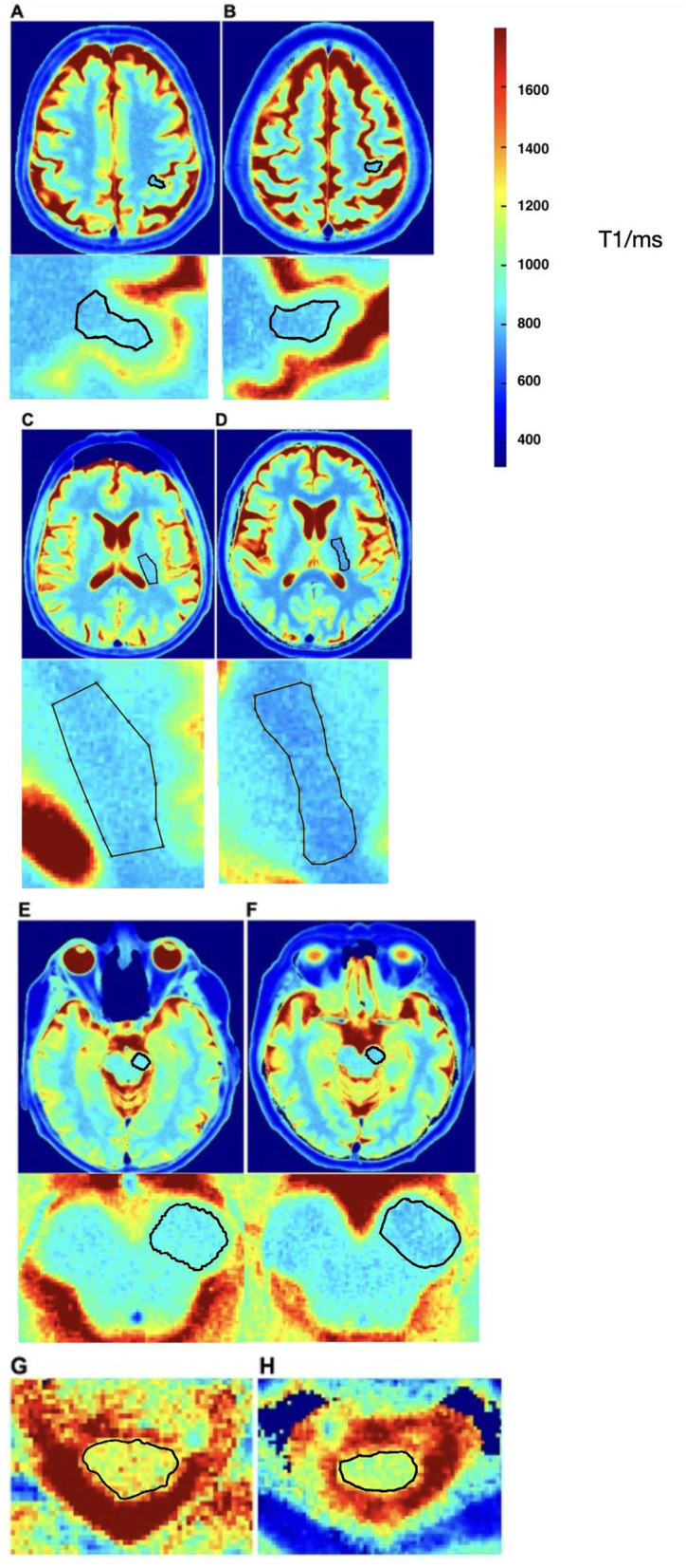
Methods: We investigated the diagnostic potential of a novel T1-mapping method based on real-time MRI with 0.5 mm in-plane resolution and 4s acquisition time per slice. The study included patients aged 18 to 90 years who met the revised El Escorial criteria for at least possible ALS. T1-relaxation times were measured along the corticospinal tract in predefined regions of interest.
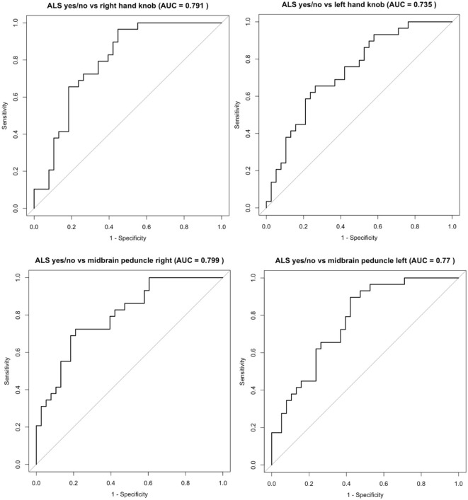
Results: Twenty-nine ALS-patients and 43 control group patients (CG) were included in the study. Median ALS Functional Rating Scale revised (ALSFRS-R) was 37 (IQR, 35-44) points and the mean duration from symptom onset to MRI was 21 ± 17 (SD) months. ALS patients showed significantly higher T1-relaxation times in all ROIs compared to CG with mean differences in the hand knob of 50 ms (p < 0.001), corona radiata 24 ms (p = 0.034), internal capsule 27 ms (p = 0.002) and midbrain peduncles 48 ms (p < 0.001). There was a consistent negative correlation between the ALSFRS-R and T1-relaxation times in all ROIs.
Conclusions: T1-relaxation times along the corticospinal tract are significantly elevated in ALS patients compared to CG and associated with lower ALSFRS-R. These results imply the analysis of T1-relaxation times as a promising diagnostic tool that can distinguish ALS patients from the control group. Ongoing longitudinal studies may provide deeper insights into disease progression and the effects of therapeutic interventions.


 分享
分享

 求助内容:
求助内容: 应助结果提醒方式:
 扫码关注我们
