Tuğba Atcı, Muhammed Burak Günay, Şirin Yaşar, Nesimi Büyükbabani, Pembegül Güneş, Şule Öztürk Sarı, Zuhal Kuş Silav, Can Baykal, Fatih Göktay
{"title":"Clinical Characteristics of Nail Unit Melanoma in Türkiye: The Experience of Two Tertiary Dermatology Centers.","authors":"Tuğba Atcı, Muhammed Burak Günay, Şirin Yaşar, Nesimi Büyükbabani, Pembegül Güneş, Şule Öztürk Sarı, Zuhal Kuş Silav, Can Baykal, Fatih Göktay","doi":"10.4274/balkanmedj.galenos.2025.2024-12-52","DOIUrl":null,"url":null,"abstract":"<p><strong>Background: </strong>The literature on the clinical presentations of nail unit melanoma (NUM) in different countries is limited.</p><p><strong>Aims: </strong>To assess the specific clinical characteristics of NUM in Türkiye.</p><p><strong>Study design: </strong>A retrospective cross sectional study.</p><p><strong>Methods: </strong>Patients with NUM in two centers were retrospectively evaluated for their clinicopathological features, including the location, laterality, destruction of the nail plate, erosion or ulceration, presence of longitudinal melanonychia (LM), Hutchinson's sign (HS), and the absence of pigmentation and Breslow thickness. These variables were compared in terms of the main location of the NUMs (fingernail versus toenail).</p><p><strong>Results: </strong>A total of 37 patients (54.1% female) of mean age 61.9 ± 14.8 years were enrolled. In most cases, NUMs were located in the fingernails (62.2%), with the most common location being the thumbnails (45.9%), followed by the big toenails (32.4%). Five cases had in situ melanoma presenting with LM. The mean Breslow thickness of invasive NUM lesions (n = 26) was 4.7 ± 4.1 mm (median: 3). Although all in situ NUMs were located on the hands, no statistically significant difference was noted in the Breslow thickness of invasive NUMs on the toenails and fingernails. NUMs were hypomelanotic/amelanotic in 10 (27%) patients. LM was clinically evident in 40.5% of the patients and was significantly more frequently observed on fingernails. The HS of the nail folds was noted in 40.5% of the patients, with the proximal (73.3%) and distal (73.3%) nail folds being most commonly involved. Total or partial destruction of the nail plate was recorded in 24.3% and 51.4% of the patients, respectively. Erosion and/or ulceration on the surface of the NUM was clinically present in most (75.7%) cases. Invasive NUMs associated with LM (n = 10) displayed partial destruction of the nail plate (n = 9), erosion and/or ulceration on the tumor surface (n = 7), and HS (n = 6).</p><p><strong>Conclusion: </strong>The clinical characteristics of patients with NUM, such as more common localization on the hands, a high rate of preference for thumbnail and big toe, and the ratio of HS, were similar to the studies reported from diverse countries. Partial destruction of the nail plate is an important clinical feature of NUM. Furthermore, LM is more frequently observed in NUMs on the fingernails.</p>","PeriodicalId":8690,"journal":{"name":"Balkan Medical Journal","volume":"42 2","pages":"157-163"},"PeriodicalIF":3.8000,"publicationDate":"2025-03-03","publicationTypes":"Journal Article","fieldsOfStudy":null,"isOpenAccess":false,"openAccessPdf":"https://www.ncbi.nlm.nih.gov/pmc/articles/PMC11881541/pdf/","citationCount":"0","resultStr":null,"platform":"Semanticscholar","paperid":null,"PeriodicalName":"Balkan Medical Journal","FirstCategoryId":"3","ListUrlMain":"https://doi.org/10.4274/balkanmedj.galenos.2025.2024-12-52","RegionNum":4,"RegionCategory":"医学","ArticlePicture":[],"TitleCN":null,"AbstractTextCN":null,"PMCID":null,"EPubDate":"","PubModel":"","JCR":"Q2","JCRName":"MEDICINE, GENERAL & INTERNAL","Score":null,"Total":0}
引用次数: 0
Abstract
Background: The literature on the clinical presentations of nail unit melanoma (NUM) in different countries is limited.
Aims: To assess the specific clinical characteristics of NUM in Türkiye.
Study design: A retrospective cross sectional study.
Methods: Patients with NUM in two centers were retrospectively evaluated for their clinicopathological features, including the location, laterality, destruction of the nail plate, erosion or ulceration, presence of longitudinal melanonychia (LM), Hutchinson's sign (HS), and the absence of pigmentation and Breslow thickness. These variables were compared in terms of the main location of the NUMs (fingernail versus toenail).
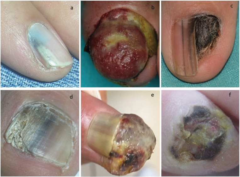
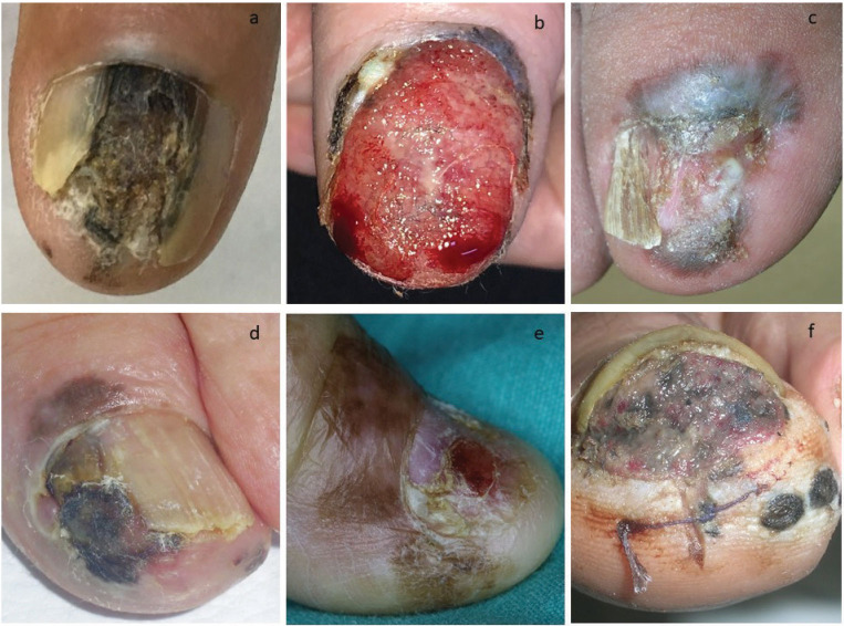
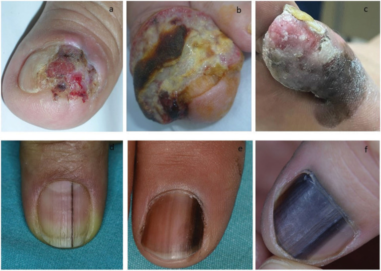
Results: A total of 37 patients (54.1% female) of mean age 61.9 ± 14.8 years were enrolled. In most cases, NUMs were located in the fingernails (62.2%), with the most common location being the thumbnails (45.9%), followed by the big toenails (32.4%). Five cases had in situ melanoma presenting with LM. The mean Breslow thickness of invasive NUM lesions (n = 26) was 4.7 ± 4.1 mm (median: 3). Although all in situ NUMs were located on the hands, no statistically significant difference was noted in the Breslow thickness of invasive NUMs on the toenails and fingernails. NUMs were hypomelanotic/amelanotic in 10 (27%) patients. LM was clinically evident in 40.5% of the patients and was significantly more frequently observed on fingernails. The HS of the nail folds was noted in 40.5% of the patients, with the proximal (73.3%) and distal (73.3%) nail folds being most commonly involved. Total or partial destruction of the nail plate was recorded in 24.3% and 51.4% of the patients, respectively. Erosion and/or ulceration on the surface of the NUM was clinically present in most (75.7%) cases. Invasive NUMs associated with LM (n = 10) displayed partial destruction of the nail plate (n = 9), erosion and/or ulceration on the tumor surface (n = 7), and HS (n = 6).
Conclusion: The clinical characteristics of patients with NUM, such as more common localization on the hands, a high rate of preference for thumbnail and big toe, and the ratio of HS, were similar to the studies reported from diverse countries. Partial destruction of the nail plate is an important clinical feature of NUM. Furthermore, LM is more frequently observed in NUMs on the fingernails.
期刊介绍:
The Balkan Medical Journal (Balkan Med J) is a peer-reviewed open-access international journal that publishes interesting clinical and experimental research conducted in all fields of medicine, interesting case reports and clinical images, invited reviews, editorials, letters, comments and letters to the Editor including reports on publication and research ethics. The journal is the official scientific publication of the Trakya University Faculty of Medicine, Edirne, Turkey and is printed six times a year, in January, March, May, July, September and November. The language of the journal is English.
The journal is based on independent and unbiased double-blinded peer-reviewed principles. Only unpublished papers that are not under review for publication elsewhere can be submitted. Balkan Medical Journal does not accept multiple submission and duplicate submission even though the previous one was published in a different language. The authors are responsible for the scientific content of the material to be published. The Balkan Medical Journal reserves the right to request any research materials on which the paper is based.
The Balkan Medical Journal encourages and enables academicians, researchers, specialists and primary care physicians of Balkan countries to publish their valuable research in all branches of medicine. The primary aim of the journal is to publish original articles with high scientific and ethical quality and serve as a good example of medical publications in the Balkans as well as in the World.


 分享
分享
 求助内容:
求助内容: 应助结果提醒方式:
 扫码关注我们
