Qi Gao, Hu Huang, Jin-Jun Shi, Ling Wang, Wei-Min Li
{"title":"Ultrasound-Guided Percutaneous Microwave Coagulation Studies on VX2 Rabbit Models for Breast Cancer Treatment and Ultrasound Imaging Assessment.","authors":"Qi Gao, Hu Huang, Jin-Jun Shi, Ling Wang, Wei-Min Li","doi":"10.2147/BCTT.S510928","DOIUrl":null,"url":null,"abstract":"<p><strong>Background: </strong>The study aimed to explore the tissue morphology and hemodynamics of rabbit VX2 breast carcinoma by high-frequency ultrasound (US) and the effectiveness and safety of US-guided percutaneous microwave coagulation (PMC) therapy on rabbit VX2 breast tumors.</p><p><strong>Methods: </strong>Twenty VX2 tumor-bearing rabbits were assessed using color Doppler ultrasound for tumor growth characteristics including echo, size, blood supply and hemodynamic parameters once a week for six weeks. Subsequently, US-guided PMC was performed in randomly assigned ten VX2 tumor-bearing rabbits (the other ten as controls). US images after ablation were obtained and analyzed. Three rabbits with double VX2 tumors were used as pathological observation at weeks 0, 1, and 4 of ablation. The therapeutic efficacy was evaluated by tumor growth, physical examinations, survival time, survival rate and metastasis of tumors and histopathology.</p><p><strong>Results: </strong>Ultrasound monitoring indicated that the tumor growth rate was 463.09% at the 2<sup>nd</sup> to 3<sup>rd</sup> weeks, and PMC was performed during this period under real-time US guidance. After microwave ablation, some tumors were greatly reduced or undetectable at week 8. Moreover, no flow signals were detected by US. The survival rates at 2 and 3 months in the treatment group and control group were 100%, 70% and 10%, 0%, respectively, while the metastatic rates were 10%, 30% and 90%, 100%, respectively <i>(P</i><0.05).</p><p><strong>Conclusion: </strong>The proliferation and metastasis of rabbit VX2 breast carcinoma were monitored by US imaging, and US-guided percutaneous microwave ablation was proven to be a safe, effective and minimally invasive therapeutic option for treating breast cancer in rabbits, showing potential clinical applicability.</p>","PeriodicalId":9106,"journal":{"name":"Breast Cancer : Targets and Therapy","volume":"17 ","pages":"241-252"},"PeriodicalIF":3.4000,"publicationDate":"2025-02-25","publicationTypes":"Journal Article","fieldsOfStudy":null,"isOpenAccess":false,"openAccessPdf":"https://www.ncbi.nlm.nih.gov/pmc/articles/PMC11871909/pdf/","citationCount":"0","resultStr":null,"platform":"Semanticscholar","paperid":null,"PeriodicalName":"Breast Cancer : Targets and Therapy","FirstCategoryId":"3","ListUrlMain":"https://doi.org/10.2147/BCTT.S510928","RegionNum":4,"RegionCategory":"医学","ArticlePicture":[],"TitleCN":null,"AbstractTextCN":null,"PMCID":null,"EPubDate":"2025/1/1 0:00:00","PubModel":"eCollection","JCR":"Q2","JCRName":"ONCOLOGY","Score":null,"Total":0}
引用次数: 0
Abstract
Background: The study aimed to explore the tissue morphology and hemodynamics of rabbit VX2 breast carcinoma by high-frequency ultrasound (US) and the effectiveness and safety of US-guided percutaneous microwave coagulation (PMC) therapy on rabbit VX2 breast tumors.
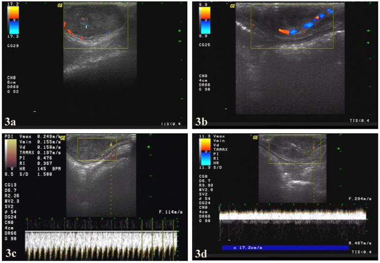
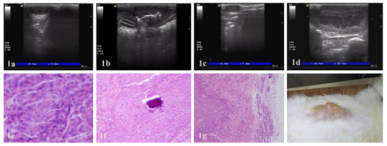
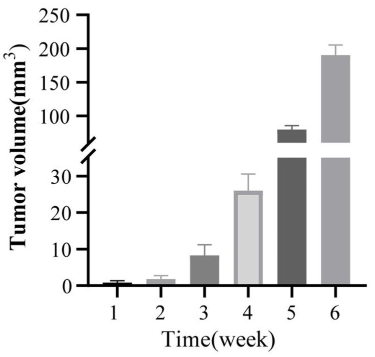
Methods: Twenty VX2 tumor-bearing rabbits were assessed using color Doppler ultrasound for tumor growth characteristics including echo, size, blood supply and hemodynamic parameters once a week for six weeks. Subsequently, US-guided PMC was performed in randomly assigned ten VX2 tumor-bearing rabbits (the other ten as controls). US images after ablation were obtained and analyzed. Three rabbits with double VX2 tumors were used as pathological observation at weeks 0, 1, and 4 of ablation. The therapeutic efficacy was evaluated by tumor growth, physical examinations, survival time, survival rate and metastasis of tumors and histopathology.
Results: Ultrasound monitoring indicated that the tumor growth rate was 463.09% at the 2nd to 3rd weeks, and PMC was performed during this period under real-time US guidance. After microwave ablation, some tumors were greatly reduced or undetectable at week 8. Moreover, no flow signals were detected by US. The survival rates at 2 and 3 months in the treatment group and control group were 100%, 70% and 10%, 0%, respectively, while the metastatic rates were 10%, 30% and 90%, 100%, respectively (P<0.05).
Conclusion: The proliferation and metastasis of rabbit VX2 breast carcinoma were monitored by US imaging, and US-guided percutaneous microwave ablation was proven to be a safe, effective and minimally invasive therapeutic option for treating breast cancer in rabbits, showing potential clinical applicability.


 分享
分享
 求助内容:
求助内容: 应助结果提醒方式:
 扫码关注我们
