{"title":"Primary Follicular Lymphoma of Thyroid: A Rare Case Report with Review of the Literature.","authors":"Shruthi K P, Lincy Joseph, Jeena V Chimmen","doi":"10.30699/ijp.2024.562997.2985","DOIUrl":null,"url":null,"abstract":"<p><strong>Background & objective: </strong>Thyroid lymphomas are predominantly secondary to lymphoma at other sites, and primary follicular lymphoma of the thyroid is a very rare entity.</p><p><strong>Case presentation: </strong>Here, we report a case of a 62-year-old female who presented with swelling in the front of her neck for one month. The clinical diagnosis was a multinodular goiter. Fine needle aspiration cytology was done and reported as nodular colloid goiter with lymphocytic thyroiditis. The system examination was unremarkable. Histopathological assessments of the right hemithyroidectomy specimen revealed the effacement of thyroid architecture by abnormal and extensive lymphoid follicles. Immunohistochemistry revealed CD20, CD10, BCL2, and BCL6 positivity in the lymphoid follicles. FDG-PT CT scan demonstrated no evidence of lymphoma elsewhere. So, a e final diagnosis of follicular lymphoma of the thyroid was made.</p><p><strong>Conclusion: </strong>Due to the rarity and low prevalence of primary follicular lymphoma of the thyroid and challenging in its differentiation from Hashimoto's thyroiditis with dense lymphoplasmacytic infiltration and formation of lymphoid follicles, histopathologic diagnosis should be confirmed by immunohistochemical studies.</p>","PeriodicalId":38900,"journal":{"name":"Iranian Journal of Pathology","volume":"19 4","pages":"447-452"},"PeriodicalIF":0.0000,"publicationDate":"2024-01-01","publicationTypes":"Journal Article","fieldsOfStudy":null,"isOpenAccess":false,"openAccessPdf":"https://www.ncbi.nlm.nih.gov/pmc/articles/PMC11872031/pdf/","citationCount":"0","resultStr":null,"platform":"Semanticscholar","paperid":null,"PeriodicalName":"Iranian Journal of Pathology","FirstCategoryId":"1085","ListUrlMain":"https://doi.org/10.30699/ijp.2024.562997.2985","RegionNum":0,"RegionCategory":null,"ArticlePicture":[],"TitleCN":null,"AbstractTextCN":null,"PMCID":null,"EPubDate":"2024/10/2 0:00:00","PubModel":"Epub","JCR":"Q3","JCRName":"Medicine","Score":null,"Total":0}
引用次数: 0
Abstract
Background & objective: Thyroid lymphomas are predominantly secondary to lymphoma at other sites, and primary follicular lymphoma of the thyroid is a very rare entity.
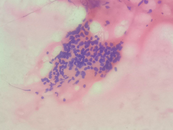
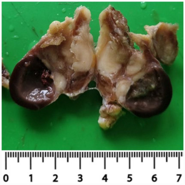
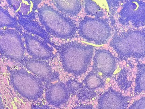
Case presentation: Here, we report a case of a 62-year-old female who presented with swelling in the front of her neck for one month. The clinical diagnosis was a multinodular goiter. Fine needle aspiration cytology was done and reported as nodular colloid goiter with lymphocytic thyroiditis. The system examination was unremarkable. Histopathological assessments of the right hemithyroidectomy specimen revealed the effacement of thyroid architecture by abnormal and extensive lymphoid follicles. Immunohistochemistry revealed CD20, CD10, BCL2, and BCL6 positivity in the lymphoid follicles. FDG-PT CT scan demonstrated no evidence of lymphoma elsewhere. So, a e final diagnosis of follicular lymphoma of the thyroid was made.
Conclusion: Due to the rarity and low prevalence of primary follicular lymphoma of the thyroid and challenging in its differentiation from Hashimoto's thyroiditis with dense lymphoplasmacytic infiltration and formation of lymphoid follicles, histopathologic diagnosis should be confirmed by immunohistochemical studies.


 分享
分享
 求助内容:
求助内容: 应助结果提醒方式:
 扫码关注我们
