Alec T Beeve, Mohamed G Hassan, Anna Li, Nicole Migotsky, Matthew J Silva, Erica L Scheller
{"title":"Spatial histomorphometry reveals that local peripheral nerves modulate but are not required for skeletal adaptation to applied load in mice.","authors":"Alec T Beeve, Mohamed G Hassan, Anna Li, Nicole Migotsky, Matthew J Silva, Erica L Scheller","doi":"10.1093/jbmrpl/ziaf006","DOIUrl":null,"url":null,"abstract":"<p><p>Mechanical loading is required for bone health and results in skeletal adaptation to optimize strength. Local nerve axons, particularly within the periosteum, may respond to load-induced biomechanical and biochemical cues. However, their role in the bone anabolic response remains controversial. We hypothesized that spatial alignment of periosteal nerves with sites of load-induced bone formation would clarify this relationship. To achieve this, we developed RadialQuant, a custom tool for spatial histomorphometry. Tibiae of control and neurectomized (sciatic/femoral nerve cut) pan-neuronal Baf53b-tdTomato reporter mice were loaded for 5 days. Bone formation and periosteal nerve axon density were then quantified simultaneously in non-decalcified sections of the mid-diaphysis using RadialQuant. In control animals, anabolic loading induced maximal periosteal bone formation at the site of peak compression, as has been reported previously. By contrast, loading did not significantly change overall periosteal nerve density. Neurectomy depleted ~90% of all periosteal axons, with near-total depletion on load-responsive surfaces. Neurectomy alone also caused de novo bone formation on the lateral aspect of the mid-diaphysis. However, neurectomy did not inhibit load-induced increases in periosteal bone area, mineralizing surface, or bone formation rate. Rather, neurectomy spatially redistributed load-induced bone formation toward the lateral tibial surface with a reduction in periosteal bone formation at the posterolateral apex (-63%) and enhancement at the lateral surface (+1360%). Altogether, this contributed to comparable load-induced changes in cortical bone area fraction. Our results show that local skeletal innervation modulates but is not required for skeletal adaptation to applied load in our model. This supports the continued use of loading and weight-bearing exercise as an effective strategy to increase bone mass, even in settings of peripheral nerve damage or dysfunction.</p>","PeriodicalId":14611,"journal":{"name":"JBMR Plus","volume":"9 3","pages":"ziaf006"},"PeriodicalIF":2.4000,"publicationDate":"2025-01-12","publicationTypes":"Journal Article","fieldsOfStudy":null,"isOpenAccess":false,"openAccessPdf":"https://www.ncbi.nlm.nih.gov/pmc/articles/PMC11878550/pdf/","citationCount":"0","resultStr":null,"platform":"Semanticscholar","paperid":null,"PeriodicalName":"JBMR Plus","FirstCategoryId":"1085","ListUrlMain":"https://doi.org/10.1093/jbmrpl/ziaf006","RegionNum":0,"RegionCategory":null,"ArticlePicture":[],"TitleCN":null,"AbstractTextCN":null,"PMCID":null,"EPubDate":"2025/3/1 0:00:00","PubModel":"eCollection","JCR":"Q2","JCRName":"ENDOCRINOLOGY & METABOLISM","Score":null,"Total":0}
引用次数: 0
Abstract
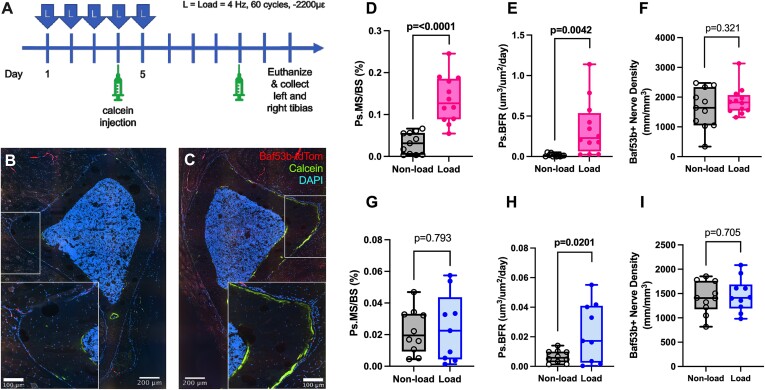
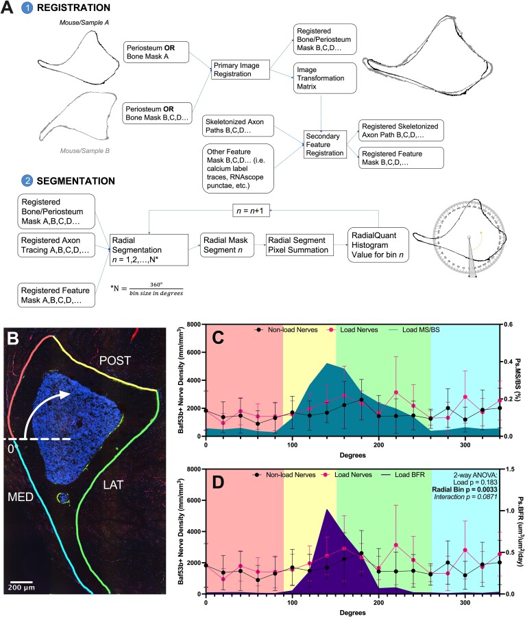
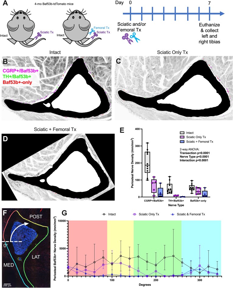
Mechanical loading is required for bone health and results in skeletal adaptation to optimize strength. Local nerve axons, particularly within the periosteum, may respond to load-induced biomechanical and biochemical cues. However, their role in the bone anabolic response remains controversial. We hypothesized that spatial alignment of periosteal nerves with sites of load-induced bone formation would clarify this relationship. To achieve this, we developed RadialQuant, a custom tool for spatial histomorphometry. Tibiae of control and neurectomized (sciatic/femoral nerve cut) pan-neuronal Baf53b-tdTomato reporter mice were loaded for 5 days. Bone formation and periosteal nerve axon density were then quantified simultaneously in non-decalcified sections of the mid-diaphysis using RadialQuant. In control animals, anabolic loading induced maximal periosteal bone formation at the site of peak compression, as has been reported previously. By contrast, loading did not significantly change overall periosteal nerve density. Neurectomy depleted ~90% of all periosteal axons, with near-total depletion on load-responsive surfaces. Neurectomy alone also caused de novo bone formation on the lateral aspect of the mid-diaphysis. However, neurectomy did not inhibit load-induced increases in periosteal bone area, mineralizing surface, or bone formation rate. Rather, neurectomy spatially redistributed load-induced bone formation toward the lateral tibial surface with a reduction in periosteal bone formation at the posterolateral apex (-63%) and enhancement at the lateral surface (+1360%). Altogether, this contributed to comparable load-induced changes in cortical bone area fraction. Our results show that local skeletal innervation modulates but is not required for skeletal adaptation to applied load in our model. This supports the continued use of loading and weight-bearing exercise as an effective strategy to increase bone mass, even in settings of peripheral nerve damage or dysfunction.


 分享
分享
 求助内容:
求助内容: 应助结果提醒方式:
 扫码关注我们
