Rosa Catalano, Emma Nozza, Barbara Altieri, Emanuela Esposito, Giorgio A Croci, Anna Maria Barbieri, Donatella Treppiedi, Sonia Di Bari, Otilia Kimpel, Mario Detomas, Mariangela Tamburello, Marc P Schauer, Sabine Herterich, Anna Angelousi, Michaela Luconi, Letizia Canu, Gabriella Nesi, Constanze Hantel, Sandra Sigala, Laura-Sophie Landwehr, Guido Di Dalmazi, Elisa Cassinotti, Ludovica Baldari, Serena Palmieri, Alessandra Mangone, Emanuele Ferrante, Cristina L Ronchi, Giovanna Mantovani, Erika Peverelli
{"title":"Emerging role of IGF1R and IR expression and localisation in adrenocortical carcinomas.","authors":"Rosa Catalano, Emma Nozza, Barbara Altieri, Emanuela Esposito, Giorgio A Croci, Anna Maria Barbieri, Donatella Treppiedi, Sonia Di Bari, Otilia Kimpel, Mario Detomas, Mariangela Tamburello, Marc P Schauer, Sabine Herterich, Anna Angelousi, Michaela Luconi, Letizia Canu, Gabriella Nesi, Constanze Hantel, Sandra Sigala, Laura-Sophie Landwehr, Guido Di Dalmazi, Elisa Cassinotti, Ludovica Baldari, Serena Palmieri, Alessandra Mangone, Emanuele Ferrante, Cristina L Ronchi, Giovanna Mantovani, Erika Peverelli","doi":"10.1186/s12964-025-02115-0","DOIUrl":null,"url":null,"abstract":"<p><strong>Background: </strong>The insulin-like growth factor 2 (IGF2) is overexpressed in 90% of adrenocortical carcinomas (ACC) and promotes cell proliferation via IGF1R and isoform A of insulin receptor (IRA). However, IGF2 role in ACC tumourigenesis has not been completely understood yet, and the contribution of IGF1R and IRA in mediating ACC cell growth has been poorly explored. This study aimed to investigate IGF1R and IR expression and localisation, including the expression of IR isoforms, in ACC and adrenocortical adenomas (ACA), and their role in IGF2-driven proliferation.</p><p><strong>Methods: </strong>Immunohistochemistry staining of IGF1R and IR was performed on 118 ACC and 22 ACA to evaluate their expression and cellular localisation and statistical analyses were carried out to assess correlations with clinicopathological data. The expression of IRA and IRB in ACC and ACA tissues, ACC cell lines and ACC and ACA primary cultures was determined by RT-qPCR. To appraise the specific role of IGF1R and IR in mediating IGF2 mitogenic pathway, single and double silencing of receptors and their inhibition in 2 ACC cell lines derived from primary tumours (H295R and JIL-2266) and 2 derived from metastatic tumours (MUC-1 and TVBF-7) as well as in ACC and ACA primary cultures were performed.</p><p><strong>Results: </strong>We found a higher IGF1R plasma membrane localisation in ACC compared to ACA. In ACC this localisation was associated with higher Ki67 and Weiss score. IR was expressed in about half of ACC and in all ACA but, in ACC, it was associated with higher Ki67 and Weiss score. RT-qPCR revealed that the prevalent isoform of IR was IRA in ACC and ACA, but not in normal adrenals. In ACC cell lines, double IGF1R + IR silencing reduced cell proliferation in JIL-2266, MUC-1 and TVBF-7 but not in H295R. In ACC, but not ACA, primary cultures, cell proliferation was reduced after IR but not IGF1R knockdown.</p><p><strong>Conclusions: </strong>Overall, these data suggest that IGF1R localisation and IR expression represent new biomarkers predicting tumour aggressiveness, as well as possible molecular markers useful to patients' stratification for more individualized IGF1R-IR targeted therapies or for novel pharmacological approaches specifically targeting IRA isoform.</p>","PeriodicalId":55268,"journal":{"name":"Cell Communication and Signaling","volume":"23 1","pages":"119"},"PeriodicalIF":8.2000,"publicationDate":"2025-03-04","publicationTypes":"Journal Article","fieldsOfStudy":null,"isOpenAccess":false,"openAccessPdf":"https://www.ncbi.nlm.nih.gov/pmc/articles/PMC11877998/pdf/","citationCount":"0","resultStr":null,"platform":"Semanticscholar","paperid":null,"PeriodicalName":"Cell Communication and Signaling","FirstCategoryId":"99","ListUrlMain":"https://doi.org/10.1186/s12964-025-02115-0","RegionNum":2,"RegionCategory":"生物学","ArticlePicture":[],"TitleCN":null,"AbstractTextCN":null,"PMCID":null,"EPubDate":"","PubModel":"","JCR":"Q1","JCRName":"CELL BIOLOGY","Score":null,"Total":0}
引用次数: 0
Abstract
Background: The insulin-like growth factor 2 (IGF2) is overexpressed in 90% of adrenocortical carcinomas (ACC) and promotes cell proliferation via IGF1R and isoform A of insulin receptor (IRA). However, IGF2 role in ACC tumourigenesis has not been completely understood yet, and the contribution of IGF1R and IRA in mediating ACC cell growth has been poorly explored. This study aimed to investigate IGF1R and IR expression and localisation, including the expression of IR isoforms, in ACC and adrenocortical adenomas (ACA), and their role in IGF2-driven proliferation.
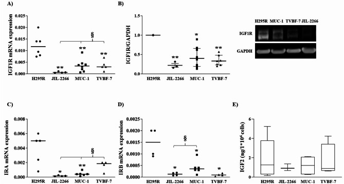
Methods: Immunohistochemistry staining of IGF1R and IR was performed on 118 ACC and 22 ACA to evaluate their expression and cellular localisation and statistical analyses were carried out to assess correlations with clinicopathological data. The expression of IRA and IRB in ACC and ACA tissues, ACC cell lines and ACC and ACA primary cultures was determined by RT-qPCR. To appraise the specific role of IGF1R and IR in mediating IGF2 mitogenic pathway, single and double silencing of receptors and their inhibition in 2 ACC cell lines derived from primary tumours (H295R and JIL-2266) and 2 derived from metastatic tumours (MUC-1 and TVBF-7) as well as in ACC and ACA primary cultures were performed.
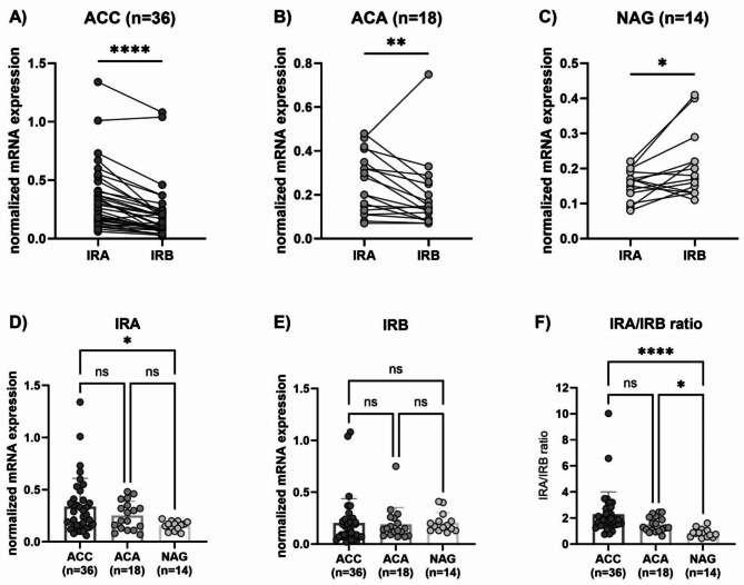
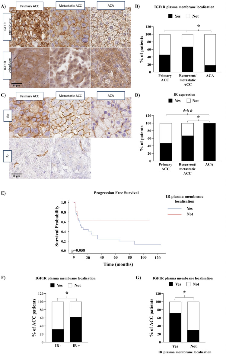
Results: We found a higher IGF1R plasma membrane localisation in ACC compared to ACA. In ACC this localisation was associated with higher Ki67 and Weiss score. IR was expressed in about half of ACC and in all ACA but, in ACC, it was associated with higher Ki67 and Weiss score. RT-qPCR revealed that the prevalent isoform of IR was IRA in ACC and ACA, but not in normal adrenals. In ACC cell lines, double IGF1R + IR silencing reduced cell proliferation in JIL-2266, MUC-1 and TVBF-7 but not in H295R. In ACC, but not ACA, primary cultures, cell proliferation was reduced after IR but not IGF1R knockdown.
Conclusions: Overall, these data suggest that IGF1R localisation and IR expression represent new biomarkers predicting tumour aggressiveness, as well as possible molecular markers useful to patients' stratification for more individualized IGF1R-IR targeted therapies or for novel pharmacological approaches specifically targeting IRA isoform.
期刊介绍:
Cell Communication and Signaling (CCS) is a peer-reviewed, open-access scientific journal that focuses on cellular signaling pathways in both normal and pathological conditions. It publishes original research, reviews, and commentaries, welcoming studies that utilize molecular, morphological, biochemical, structural, and cell biology approaches. CCS also encourages interdisciplinary work and innovative models, including in silico, in vitro, and in vivo approaches, to facilitate investigations of cell signaling pathways, networks, and behavior.
Starting from January 2019, CCS is proud to announce its affiliation with the International Cell Death Society. The journal now encourages submissions covering all aspects of cell death, including apoptotic and non-apoptotic mechanisms, cell death in model systems, autophagy, clearance of dying cells, and the immunological and pathological consequences of dying cells in the tissue microenvironment.


 分享
分享
 求助内容:
求助内容: 应助结果提醒方式:
 扫码关注我们
