1α,25-Dihydroxyvitamin D3 accelerates skin wound re-epithelialization by promoting epidermal stem cell proliferation and differentiation through PI3K activation: an in vitro and in vivo study.
{"title":"1α,25-Dihydroxyvitamin D3 accelerates skin wound re-epithelialization by promoting epidermal stem cell proliferation and differentiation through PI3K activation: an in vitro and in vivo study.","authors":"Rongshuai Yan, Zhihui Liu, Song Wang, Dongli Fan","doi":"10.1590/1414-431X2025e14121","DOIUrl":null,"url":null,"abstract":"<p><p>1α,25-Dihydroxyvitamin D3 (VD3), the active form of vitamin D, plays a crucial role in wound healing. In this study, we aimed to investigate the effect of VD3 on the proliferation and differentiation of epidermal stem cells (EpSCs) and monitor its impact on re-epithelialization. We established a murine full-thickness skin defect model and applied four doses of VD3 (0, 5, 50, and 250 ng/mouse/day) to the wounds topically for three days. Immunostaining and flow cytometry confirmed the effect of VD3 on the proliferation and differentiation of EpSCs in wounds. This effect of VD3 (0, 1, 10, and 50 nM) on EpSCs and its possible mechanism were further confirmed in vitro by CCK8, westen blot, immunostaining, and flow cytometry. We found that on day five post-wounding, the means±SD length of the neo-epidermis was 195.88±11.57, 231.84±16.45, 385.80±17.50, and 268.00±8.22 μm in the control, 5, 50, and 250 ng groups, respectively, with a significant difference from the control (all P<0.05). Immunostaining and flow cytometry showed that VD3 improved the proliferation and differentiation of K15+ EpSC (vs control, all P<0.05), K14+ epidermal progenitor cells (vs control, all P<0.05), and K10+ epidermal terminal cells (vs control, all P<0.05) in vivo and in vitro. The PI3K signaling pathway appeared to underlie this response because significant inhibition of the response was found when inhibitors were used to inhibit PI3K. Our study demonstrated that VD3 is a potent promoter of cutaneous wound healing by stimulating EpSC proliferation and differentiation through PI3K activation.</p>","PeriodicalId":9088,"journal":{"name":"Brazilian Journal of Medical and Biological Research","volume":"58 ","pages":"e14121"},"PeriodicalIF":1.9000,"publicationDate":"2025-03-03","publicationTypes":"Journal Article","fieldsOfStudy":null,"isOpenAccess":false,"openAccessPdf":"https://www.ncbi.nlm.nih.gov/pmc/articles/PMC11884782/pdf/","citationCount":"0","resultStr":null,"platform":"Semanticscholar","paperid":null,"PeriodicalName":"Brazilian Journal of Medical and Biological Research","FirstCategoryId":"3","ListUrlMain":"https://doi.org/10.1590/1414-431X2025e14121","RegionNum":4,"RegionCategory":"医学","ArticlePicture":[],"TitleCN":null,"AbstractTextCN":null,"PMCID":null,"EPubDate":"2025/1/1 0:00:00","PubModel":"eCollection","JCR":"Q2","JCRName":"BIOLOGY","Score":null,"Total":0}
引用次数: 0
Abstract
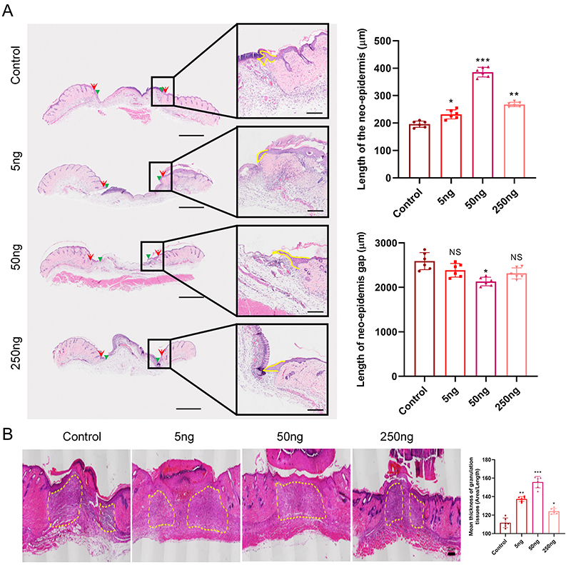
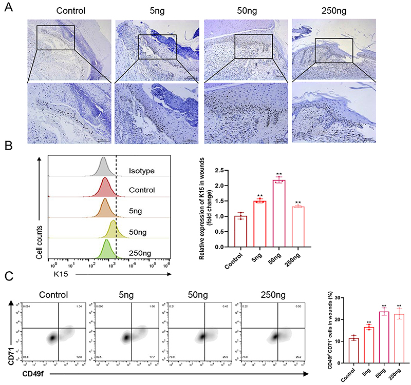
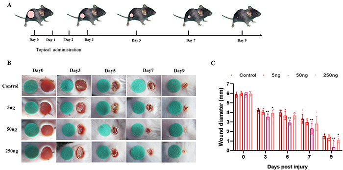
1α,25-Dihydroxyvitamin D3 (VD3), the active form of vitamin D, plays a crucial role in wound healing. In this study, we aimed to investigate the effect of VD3 on the proliferation and differentiation of epidermal stem cells (EpSCs) and monitor its impact on re-epithelialization. We established a murine full-thickness skin defect model and applied four doses of VD3 (0, 5, 50, and 250 ng/mouse/day) to the wounds topically for three days. Immunostaining and flow cytometry confirmed the effect of VD3 on the proliferation and differentiation of EpSCs in wounds. This effect of VD3 (0, 1, 10, and 50 nM) on EpSCs and its possible mechanism were further confirmed in vitro by CCK8, westen blot, immunostaining, and flow cytometry. We found that on day five post-wounding, the means±SD length of the neo-epidermis was 195.88±11.57, 231.84±16.45, 385.80±17.50, and 268.00±8.22 μm in the control, 5, 50, and 250 ng groups, respectively, with a significant difference from the control (all P<0.05). Immunostaining and flow cytometry showed that VD3 improved the proliferation and differentiation of K15+ EpSC (vs control, all P<0.05), K14+ epidermal progenitor cells (vs control, all P<0.05), and K10+ epidermal terminal cells (vs control, all P<0.05) in vivo and in vitro. The PI3K signaling pathway appeared to underlie this response because significant inhibition of the response was found when inhibitors were used to inhibit PI3K. Our study demonstrated that VD3 is a potent promoter of cutaneous wound healing by stimulating EpSC proliferation and differentiation through PI3K activation.
期刊介绍:
The Brazilian Journal of Medical and Biological Research, founded by Michel Jamra, is edited and published monthly by the Associação Brasileira de Divulgação Científica (ABDC), a federation of Brazilian scientific societies:
- Sociedade Brasileira de Biofísica (SBBf)
- Sociedade Brasileira de Farmacologia e Terapêutica Experimental (SBFTE)
- Sociedade Brasileira de Fisiologia (SBFis)
- Sociedade Brasileira de Imunologia (SBI)
- Sociedade Brasileira de Investigação Clínica (SBIC)
- Sociedade Brasileira de Neurociências e Comportamento (SBNeC).


 分享
分享
 求助内容:
求助内容: 应助结果提醒方式:
 扫码关注我们
