{"title":"Fluorescent Microscopy: A Useful and Adjunct Tool in Leprosy Diagnosis: A Cross Sectional Study.","authors":"Swati Soni, Vaishali Walke, Dinesh Asati, Anand Maurya, Sramana Mukhopadhyay","doi":"10.30699/ijp.2024.2023590.3263","DOIUrl":null,"url":null,"abstract":"<p><strong>Background & objective: </strong>Leprosy is a chronic infectious disease caused by Mycobacterium leprae. Fite-Faraco (FF) is the routine staining method used to demonstrate the presence of <i>Mycobacterium leprae</i> in tissue sections. Fluorescent microscopy (FM) can help visualize lepra bacilli better. The present study compares two methodologies, fluorescent microscopy, and Fite-Faraco, in detecting Mycobacterium leprae in tissue sections.</p><p><strong>Methods: </strong>Histopathology of skin biopsies in 60 cases of Hansen's were evaluated with FF stain. The performance of Auramine- Rhodamine Fluroscencent stain was compared with conventional FF staining in identifying Lepra bacilli.</p><p><strong>Results: </strong>A total of 60 clinically and histopathologically confirmed cases of Hansen's disease were included in this ambispective study. The cases were sub-classified into various histological categories. Auramine-rhodamine fluorescent staining was performed and examined under a fluorescent microscope with an LED light illuminator. The bacteriological index (BI) was calculated under an oil immersion field for both Fite-Faraco (FF) staining and fluorescent microscopy (FM), graded from zero to six plus according to Ridley's logarithmic scale. Lepra bacilli were identified in 70% of patients on FF staining, while fluorescent microscopy showed positivity in 80%. The mean BI calculated by FM (2.48) was significantly higher than that by the FF method (2.18), and more multibacillary disease was identified by fluorescent staining compared to FF staining.</p><p><strong>Conclusion: </strong>It is advantageous to use fluorescent microscopy as an adjunct to conventional Fite-Faraco stain especially in cases where the latter fails to detect lepra bacilli and in a clinically suspected multibacillary disease.</p>","PeriodicalId":38900,"journal":{"name":"Iranian Journal of Pathology","volume":"20 1","pages":"33-41"},"PeriodicalIF":0.0000,"publicationDate":"2025-01-01","publicationTypes":"Journal Article","fieldsOfStudy":null,"isOpenAccess":false,"openAccessPdf":"https://www.ncbi.nlm.nih.gov/pmc/articles/PMC11887632/pdf/","citationCount":"0","resultStr":null,"platform":"Semanticscholar","paperid":null,"PeriodicalName":"Iranian Journal of Pathology","FirstCategoryId":"1085","ListUrlMain":"https://doi.org/10.30699/ijp.2024.2023590.3263","RegionNum":0,"RegionCategory":null,"ArticlePicture":[],"TitleCN":null,"AbstractTextCN":null,"PMCID":null,"EPubDate":"2025/1/10 0:00:00","PubModel":"Epub","JCR":"Q3","JCRName":"Medicine","Score":null,"Total":0}
引用次数: 0
Abstract
Background & objective: Leprosy is a chronic infectious disease caused by Mycobacterium leprae. Fite-Faraco (FF) is the routine staining method used to demonstrate the presence of Mycobacterium leprae in tissue sections. Fluorescent microscopy (FM) can help visualize lepra bacilli better. The present study compares two methodologies, fluorescent microscopy, and Fite-Faraco, in detecting Mycobacterium leprae in tissue sections.
Methods: Histopathology of skin biopsies in 60 cases of Hansen's were evaluated with FF stain. The performance of Auramine- Rhodamine Fluroscencent stain was compared with conventional FF staining in identifying Lepra bacilli.
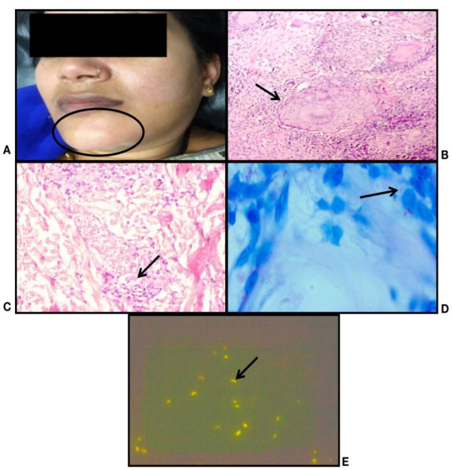
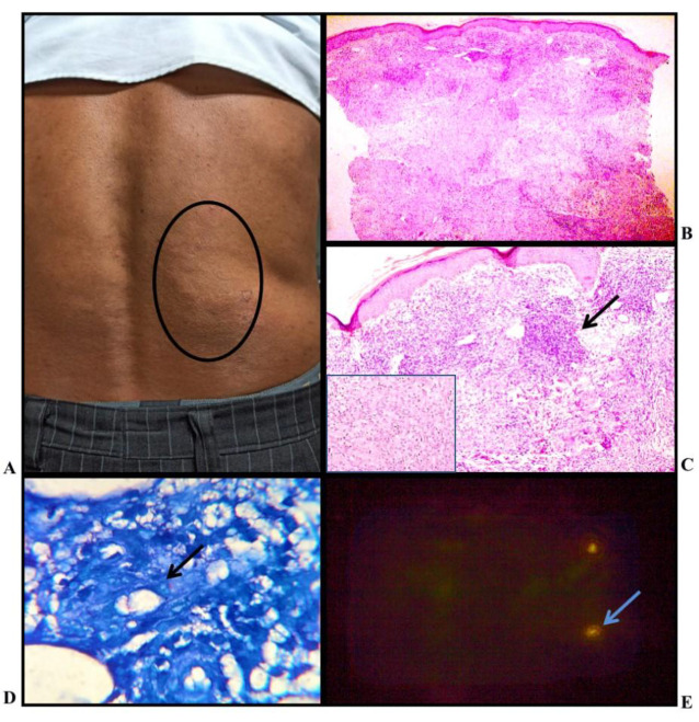
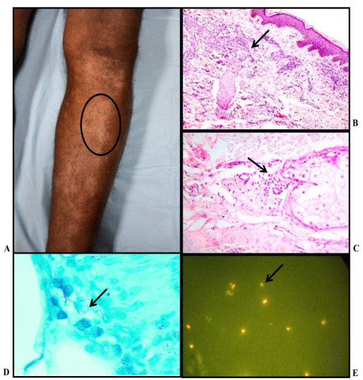
Results: A total of 60 clinically and histopathologically confirmed cases of Hansen's disease were included in this ambispective study. The cases were sub-classified into various histological categories. Auramine-rhodamine fluorescent staining was performed and examined under a fluorescent microscope with an LED light illuminator. The bacteriological index (BI) was calculated under an oil immersion field for both Fite-Faraco (FF) staining and fluorescent microscopy (FM), graded from zero to six plus according to Ridley's logarithmic scale. Lepra bacilli were identified in 70% of patients on FF staining, while fluorescent microscopy showed positivity in 80%. The mean BI calculated by FM (2.48) was significantly higher than that by the FF method (2.18), and more multibacillary disease was identified by fluorescent staining compared to FF staining.
Conclusion: It is advantageous to use fluorescent microscopy as an adjunct to conventional Fite-Faraco stain especially in cases where the latter fails to detect lepra bacilli and in a clinically suspected multibacillary disease.


 分享
分享
 求助内容:
求助内容: 应助结果提醒方式:
 扫码关注我们
