{"title":"Equilibrium phase images of the liver using a contrast-enhancement boost instead of the portal vein phase.","authors":"Yuji Tachibana, Kenichiro Otsuka, Tomoaki Shiroo, Yoshiki Asayama","doi":"10.4329/wjr.v17.i2.102462","DOIUrl":null,"url":null,"abstract":"<p><strong>Background: </strong>Three-phase dynamic computed tomography imaging is particularly useful in the liver region. However, dynamic imaging with contrast media has the disadvantage of increased radiation exposure due to multiple imaging sessions. We hypothesized that the contrast enhancement boost (CE-boost) technique could be used to enhance the contrast in equilibrium phase (EP) images and produce enhancement similar to that of portal vein phase (PVP) images, and if this is possible, EP imaging could play the same role as PVP imaging. We also speculated that this might allow the conversion of three-phase dynamic imaging to biphasic dynamic imaging, reducing patients' radiation exposure.</p><p><strong>Aim: </strong>To determine if the CE-boost of EP, CE-boost (EP) is useful compared to a conventional image.</p><p><strong>Methods: </strong>We retrospectively analyzed the cases of 52 patients who were diagnosed with liver cancer between January 2016 and October 2022 at our institution. From these computed tomography images, CE-boost images were generated from the EP and plane images. We compared the PVP, EP, and CE-boost (EP) for blood vessels and hepatic parenchyma based on the contrast-to-noise ratio (CNR), signal-to-noise ratio, and figure-of-merit (FOM). Visual assessments were also performed for vessel visualization, lesion conspicuity, and image noise.</p><p><strong>Results: </strong>The CE-boost (EP) images showed significant superiority compared to the PVP images in the CNR, signal-to-noise ratio, and FOM except regarding the hepatic parenchyma. No significant differences were detected in CNR or FOM comparisons within the hepatic parenchyma (<i>P</i> = 0.62, 0.67). The comparison of the EP and CE-boost (EP) images consistently favored CE-boost (EP). Regarding the visual assessment, the CE-boost (EP) images were significantly superior to the PVP images in lesion conspicuity, and the PVP in image noise. The CE-boost (EP) images were significantly better than the EP images in the vessel visualization of segmental branches of the portal vein and lesion conspicuity, and the EP in image noise.</p><p><strong>Conclusion: </strong>The image quality of CE-boost (EP) images was comparable or superior to that of conventional PVP and EP. CE-boost (EP) images might provide information comparable to the conventional PVP.</p>","PeriodicalId":23819,"journal":{"name":"World journal of radiology","volume":"17 2","pages":"102462"},"PeriodicalIF":1.5000,"publicationDate":"2025-02-28","publicationTypes":"Journal Article","fieldsOfStudy":null,"isOpenAccess":false,"openAccessPdf":"https://www.ncbi.nlm.nih.gov/pmc/articles/PMC11885925/pdf/","citationCount":"0","resultStr":null,"platform":"Semanticscholar","paperid":null,"PeriodicalName":"World journal of radiology","FirstCategoryId":"1085","ListUrlMain":"https://doi.org/10.4329/wjr.v17.i2.102462","RegionNum":0,"RegionCategory":null,"ArticlePicture":[],"TitleCN":null,"AbstractTextCN":null,"PMCID":null,"EPubDate":"","PubModel":"","JCR":"Q3","JCRName":"RADIOLOGY, NUCLEAR MEDICINE & MEDICAL IMAGING","Score":null,"Total":0}
引用次数: 0
Abstract
Background: Three-phase dynamic computed tomography imaging is particularly useful in the liver region. However, dynamic imaging with contrast media has the disadvantage of increased radiation exposure due to multiple imaging sessions. We hypothesized that the contrast enhancement boost (CE-boost) technique could be used to enhance the contrast in equilibrium phase (EP) images and produce enhancement similar to that of portal vein phase (PVP) images, and if this is possible, EP imaging could play the same role as PVP imaging. We also speculated that this might allow the conversion of three-phase dynamic imaging to biphasic dynamic imaging, reducing patients' radiation exposure.
Aim: To determine if the CE-boost of EP, CE-boost (EP) is useful compared to a conventional image.
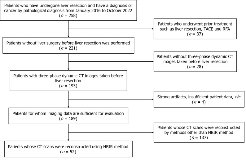
Methods: We retrospectively analyzed the cases of 52 patients who were diagnosed with liver cancer between January 2016 and October 2022 at our institution. From these computed tomography images, CE-boost images were generated from the EP and plane images. We compared the PVP, EP, and CE-boost (EP) for blood vessels and hepatic parenchyma based on the contrast-to-noise ratio (CNR), signal-to-noise ratio, and figure-of-merit (FOM). Visual assessments were also performed for vessel visualization, lesion conspicuity, and image noise.
Results: The CE-boost (EP) images showed significant superiority compared to the PVP images in the CNR, signal-to-noise ratio, and FOM except regarding the hepatic parenchyma. No significant differences were detected in CNR or FOM comparisons within the hepatic parenchyma (P = 0.62, 0.67). The comparison of the EP and CE-boost (EP) images consistently favored CE-boost (EP). Regarding the visual assessment, the CE-boost (EP) images were significantly superior to the PVP images in lesion conspicuity, and the PVP in image noise. The CE-boost (EP) images were significantly better than the EP images in the vessel visualization of segmental branches of the portal vein and lesion conspicuity, and the EP in image noise.
Conclusion: The image quality of CE-boost (EP) images was comparable or superior to that of conventional PVP and EP. CE-boost (EP) images might provide information comparable to the conventional PVP.


 分享
分享
 求助内容:
求助内容: 应助结果提醒方式:
 扫码关注我们
