{"title":"Vision Loss in Neurocysticercosis: A Systematic Review of Case Reports and Series.","authors":"Ravindra Kumar Garg, Pragati Garg, Vimal Kumar Paliwal, Shweta Pandey","doi":"10.3988/jcn.2024.0565","DOIUrl":null,"url":null,"abstract":"<p><strong>Background and purpose: </strong>Neurocysticercosis is a parasitic infection caused by <i>Taenia solium</i> larvae that leads to various neurological symptoms, including vision loss. This systematic review analyzed cases of vision loss associated with neurocysticercosis to assess its etiology and vision outcomes.</p><p><strong>Methods: </strong>Following PRISMA guidelines, the review included reports on human subjects with vision loss due to neurocysticercosis and is registered with PROSPERO (CRD42024556278). The PubMed, Scopus, Embase, and Google Scholar databases were searched.</p><p><strong>Results: </strong>This review included 149 records from 176 patients with a mean age of 27.5 years, comprising 40.3% females, 59.1% males, and 0.6% subjects of unknown sex. Most cases were from Asia, predominantly India. The illness duration varied, but was mostly between 1 and 6 months. In addition to vision loss, common symptoms were headache or orbital pain (30.7%), seizures (12.5%), and altered consciousness (5.7%). Vision loss was mainly unilateral (72.7%). Imaging abnormalities included multiple cystic brain lesions (16.5%), enhanced lesions (4.0%), and calcified lesions (2.3%). Intravitreal and retinal regions were most affected (52.3%), followed by the anterior chamber (6.2%), orbital apex (5.1%), and optic nerve (6.2%). Anticysticercal drugs were the primary treatment, with 57.4% of cases showing improvement. Surgical excision was performed in 40.9% of cases with intravitreal or retinal cysts.</p><p><strong>Conclusions: </strong>Vision loss in neurocysticercosis is mainly due to intravitreal and retinal involvement, and is frequently associated with multiple cystic brain lesions. Anticysticercal drugs can produce improvements, though surgical intervention is often needed for intravitreal or retinal cysts. Most of the patients in this review improved, though severe outcomes such as eye loss were reported.</p>","PeriodicalId":15432,"journal":{"name":"Journal of Clinical Neurology","volume":"21 2","pages":"137-145"},"PeriodicalIF":3.1000,"publicationDate":"2025-03-01","publicationTypes":"Journal Article","fieldsOfStudy":null,"isOpenAccess":false,"openAccessPdf":"https://www.ncbi.nlm.nih.gov/pmc/articles/PMC11896748/pdf/","citationCount":"0","resultStr":null,"platform":"Semanticscholar","paperid":null,"PeriodicalName":"Journal of Clinical Neurology","FirstCategoryId":"3","ListUrlMain":"https://doi.org/10.3988/jcn.2024.0565","RegionNum":3,"RegionCategory":"医学","ArticlePicture":[],"TitleCN":null,"AbstractTextCN":null,"PMCID":null,"EPubDate":"","PubModel":"","JCR":"Q2","JCRName":"CLINICAL NEUROLOGY","Score":null,"Total":0}
引用次数: 0
Abstract
Background and purpose: Neurocysticercosis is a parasitic infection caused by Taenia solium larvae that leads to various neurological symptoms, including vision loss. This systematic review analyzed cases of vision loss associated with neurocysticercosis to assess its etiology and vision outcomes.
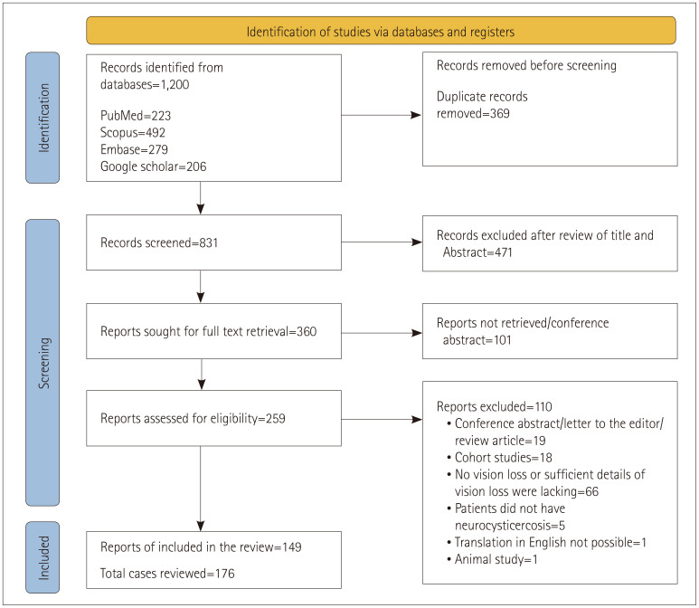
Methods: Following PRISMA guidelines, the review included reports on human subjects with vision loss due to neurocysticercosis and is registered with PROSPERO (CRD42024556278). The PubMed, Scopus, Embase, and Google Scholar databases were searched.
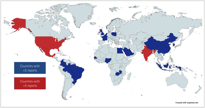
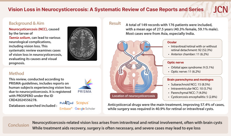
Results: This review included 149 records from 176 patients with a mean age of 27.5 years, comprising 40.3% females, 59.1% males, and 0.6% subjects of unknown sex. Most cases were from Asia, predominantly India. The illness duration varied, but was mostly between 1 and 6 months. In addition to vision loss, common symptoms were headache or orbital pain (30.7%), seizures (12.5%), and altered consciousness (5.7%). Vision loss was mainly unilateral (72.7%). Imaging abnormalities included multiple cystic brain lesions (16.5%), enhanced lesions (4.0%), and calcified lesions (2.3%). Intravitreal and retinal regions were most affected (52.3%), followed by the anterior chamber (6.2%), orbital apex (5.1%), and optic nerve (6.2%). Anticysticercal drugs were the primary treatment, with 57.4% of cases showing improvement. Surgical excision was performed in 40.9% of cases with intravitreal or retinal cysts.
Conclusions: Vision loss in neurocysticercosis is mainly due to intravitreal and retinal involvement, and is frequently associated with multiple cystic brain lesions. Anticysticercal drugs can produce improvements, though surgical intervention is often needed for intravitreal or retinal cysts. Most of the patients in this review improved, though severe outcomes such as eye loss were reported.
期刊介绍:
The JCN aims to publish the cutting-edge research from around the world. The JCN covers clinical and translational research for physicians and researchers in the field of neurology. Encompassing the entire neurological diseases, our main focus is on the common disorders including stroke, epilepsy, Parkinson''s disease, dementia, multiple sclerosis, headache, and peripheral neuropathy. Any authors affiliated with an accredited biomedical institution may submit manuscripts of original articles, review articles, and letters to the editor. The JCN will allow clinical neurologists to enrich their knowledge of patient management, education, and clinical or experimental research, and hence their professionalism.


 分享
分享
 求助内容:
求助内容: 应助结果提醒方式:
 扫码关注我们
