Jadwiga Kleinrok, Krzysztof Kleinrok, Tadeusz Jan Popiela
{"title":"Split cord malformation - a simple, current classification based on CT and MRI neuroimaging studies.","authors":"Jadwiga Kleinrok, Krzysztof Kleinrok, Tadeusz Jan Popiela","doi":"10.5114/pjr/199683","DOIUrl":null,"url":null,"abstract":"<p><p>The aim of this paper is to present the currently used classification of split cord malformation. Split cord malformation (SCM) is a developmental defect arising during neurulation, resulting in abnormal neural tube development, with the formation of a division within the spinal cord and dural sac. The terms diastematomyelia and diplomyelia are used in the literature to describe this defect. In 1992, Pang proposed the term SCM to describe all dysraphic spinal cord defects and classified them into type I and type II, depending on the nature of the sagittal septum within the spinal canal and the presence or absence of a divided dural sac. SCM type I includes cases with a bony septum and a divided dural sac, while SCM type II includes cases without a divided dural sac but with a fibrous septum present. Depending on the type of defect, and the location and extent of the split, the condition is accompanied by neurological symptoms of varying localisation and severity. As symptoms may worsen with the child's growth, surgical intervention to remove the septum is usually necessary. In this article, the authors present the defect based on literature data, describe the current terminology regarding the defect and associated anomalies, and present a set of features that should be assessed to classify lesions.</p>","PeriodicalId":94174,"journal":{"name":"Polish journal of radiology","volume":"90 ","pages":"e46-e54"},"PeriodicalIF":0.0000,"publicationDate":"2025-01-30","publicationTypes":"Journal Article","fieldsOfStudy":null,"isOpenAccess":false,"openAccessPdf":"https://www.ncbi.nlm.nih.gov/pmc/articles/PMC11891551/pdf/","citationCount":"0","resultStr":null,"platform":"Semanticscholar","paperid":null,"PeriodicalName":"Polish journal of radiology","FirstCategoryId":"1085","ListUrlMain":"https://doi.org/10.5114/pjr/199683","RegionNum":0,"RegionCategory":null,"ArticlePicture":[],"TitleCN":null,"AbstractTextCN":null,"PMCID":null,"EPubDate":"2025/1/1 0:00:00","PubModel":"eCollection","JCR":"","JCRName":"","Score":null,"Total":0}
引用次数: 0
Abstract
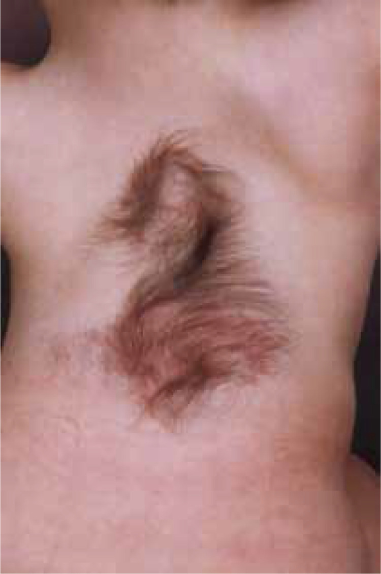
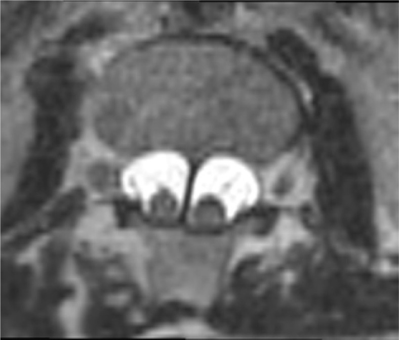
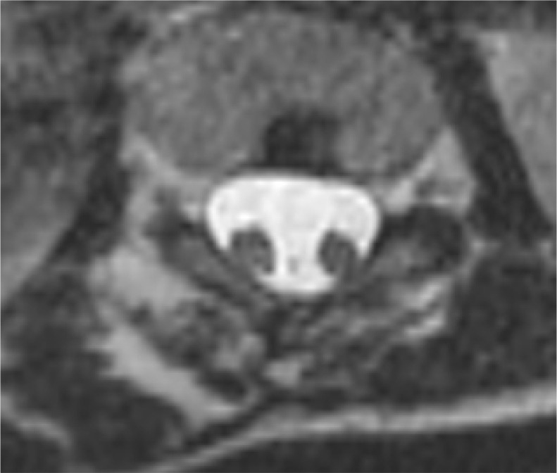
The aim of this paper is to present the currently used classification of split cord malformation. Split cord malformation (SCM) is a developmental defect arising during neurulation, resulting in abnormal neural tube development, with the formation of a division within the spinal cord and dural sac. The terms diastematomyelia and diplomyelia are used in the literature to describe this defect. In 1992, Pang proposed the term SCM to describe all dysraphic spinal cord defects and classified them into type I and type II, depending on the nature of the sagittal septum within the spinal canal and the presence or absence of a divided dural sac. SCM type I includes cases with a bony septum and a divided dural sac, while SCM type II includes cases without a divided dural sac but with a fibrous septum present. Depending on the type of defect, and the location and extent of the split, the condition is accompanied by neurological symptoms of varying localisation and severity. As symptoms may worsen with the child's growth, surgical intervention to remove the septum is usually necessary. In this article, the authors present the defect based on literature data, describe the current terminology regarding the defect and associated anomalies, and present a set of features that should be assessed to classify lesions.


 分享
分享

 求助内容:
求助内容: 应助结果提醒方式:
 扫码关注我们
