{"title":"Fallopian fimbriae entrapped in an ovarian endometriotic cyst mimicking malignancy: a case report","authors":"Atsushi Yoshida, Shigeshi Kohno, Shojiro Oka, Yuko Someya, Shigeki Arizono, Tsuyoshi Suga, Reiichi Ishikura, Hiroe Itami, Shinichiro Maeda, Kumiko Ando","doi":"10.1007/s00261-025-04882-3","DOIUrl":null,"url":null,"abstract":"<div><p>Ovarian endometriotic cysts are associated with an increased risk of clear cell and endometrioid carcinomas, as well as borderline neoplasms. Although contrast-enhancing nodules on magnetic resonance imaging (MRI) suggest malignancy, benign endometriotic cysts can also present with such features, complicating differentiation from malignancy. When malignancy is suspected, minimally invasive procedures, such as laparoscopic cystectomy, are typically avoided. However, preserving fertility and ovarian function warrants careful consideration when selecting invasive surgical procedures. From the perspective of selecting appropriate surgical approaches, accurate preoperative differentiation between benign and malignant ovarian tumors is essential. We present the first case of MRI showing fallopian fimbriae entrapped in an endometriotic cyst mimicking malignancy. A 49-year-old female presented with atypical genital bleeding. MRI revealed a right ovarian endometriotic cyst with a contrast-enhancing mural nodule (10 mm), suggestive of malignancy. The nodule demonstrated T2-weighted hypointensity equivalent to the cyst fluid without diffusion restriction. Laparotomy revealed the nodule as entrapped fallopian fimbriae within the endometriotic cyst, with no malignancy detected. In this case, the fallopian fimbriae entrapped in the endometriotic cyst appeared as an enhancing nodule because of their vascularity, mimicking malignancy. Fallopian fimbriae are inconspicuous structures that can produce false findings suggestive of malignancy, similar to other benign enhancing nodules, such as polypoid endometriosis and decidualization. However, their lack of diffusion restriction and low T2-weighted signal intensity may help distinguish them from malignancy. This knowledge is crucial for accurate diagnosis and avoiding unnecessary interventions.</p><h3>Graphical abstract</h3><div><figure><div><div><picture><source><img></source></picture></div></div></figure></div></div>","PeriodicalId":7126,"journal":{"name":"Abdominal Radiology","volume":"50 9","pages":"4374 - 4379"},"PeriodicalIF":2.2000,"publicationDate":"2025-03-17","publicationTypes":"Journal Article","fieldsOfStudy":null,"isOpenAccess":false,"openAccessPdf":"","citationCount":"0","resultStr":null,"platform":"Semanticscholar","paperid":null,"PeriodicalName":"Abdominal Radiology","FirstCategoryId":"3","ListUrlMain":"https://link.springer.com/article/10.1007/s00261-025-04882-3","RegionNum":3,"RegionCategory":"医学","ArticlePicture":[],"TitleCN":null,"AbstractTextCN":null,"PMCID":null,"EPubDate":"","PubModel":"","JCR":"Q2","JCRName":"RADIOLOGY, NUCLEAR MEDICINE & MEDICAL IMAGING","Score":null,"Total":0}
引用次数: 0
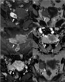
Abstract
Ovarian endometriotic cysts are associated with an increased risk of clear cell and endometrioid carcinomas, as well as borderline neoplasms. Although contrast-enhancing nodules on magnetic resonance imaging (MRI) suggest malignancy, benign endometriotic cysts can also present with such features, complicating differentiation from malignancy. When malignancy is suspected, minimally invasive procedures, such as laparoscopic cystectomy, are typically avoided. However, preserving fertility and ovarian function warrants careful consideration when selecting invasive surgical procedures. From the perspective of selecting appropriate surgical approaches, accurate preoperative differentiation between benign and malignant ovarian tumors is essential. We present the first case of MRI showing fallopian fimbriae entrapped in an endometriotic cyst mimicking malignancy. A 49-year-old female presented with atypical genital bleeding. MRI revealed a right ovarian endometriotic cyst with a contrast-enhancing mural nodule (10 mm), suggestive of malignancy. The nodule demonstrated T2-weighted hypointensity equivalent to the cyst fluid without diffusion restriction. Laparotomy revealed the nodule as entrapped fallopian fimbriae within the endometriotic cyst, with no malignancy detected. In this case, the fallopian fimbriae entrapped in the endometriotic cyst appeared as an enhancing nodule because of their vascularity, mimicking malignancy. Fallopian fimbriae are inconspicuous structures that can produce false findings suggestive of malignancy, similar to other benign enhancing nodules, such as polypoid endometriosis and decidualization. However, their lack of diffusion restriction and low T2-weighted signal intensity may help distinguish them from malignancy. This knowledge is crucial for accurate diagnosis and avoiding unnecessary interventions.
期刊介绍:
Abdominal Radiology seeks to meet the professional needs of the abdominal radiologist by publishing clinically pertinent original, review and practice related articles on the gastrointestinal and genitourinary tracts and abdominal interventional and radiologic procedures. Case reports are generally not accepted unless they are the first report of a new disease or condition, or part of a special solicited section.
Reasons to Publish Your Article in Abdominal Radiology:
· Official journal of the Society of Abdominal Radiology (SAR)
· Published in Cooperation with:
European Society of Gastrointestinal and Abdominal Radiology (ESGAR)
European Society of Urogenital Radiology (ESUR)
Asian Society of Abdominal Radiology (ASAR)
· Efficient handling and Expeditious review
· Author feedback is provided in a mentoring style
· Global readership
· Readers can earn CME credits


 分享
分享
 求助内容:
求助内容: 应助结果提醒方式:
 扫码关注我们
