Andres Server, Anna Latysheva, Bård Nedregaard, Arild Erland Rønnestad, Pål Bache Marthinsen
{"title":"Neonatal subpial hemorrhage: clinical presentation, neuroimaging findings and outcome.","authors":"Andres Server, Anna Latysheva, Bård Nedregaard, Arild Erland Rønnestad, Pål Bache Marthinsen","doi":"10.1007/s00234-025-03589-y","DOIUrl":null,"url":null,"abstract":"<p><strong>Purpose: </strong>Subpial hemorrhage is a rare form of intracranial hemorrhage (ICH) in neonates that remains underreported and inadequately understood. The aim of this study is to characterize the neuroimaging patterns of subpial hemorrhage, assess changes in the underlying brain parenchyma, and examine its clinical features and outcomes.</p><p><strong>Methods: </strong>We reviewed the medical records and neuroimaging data of neonates with subpial hemorrhage admitted to our hospital between January 2010 and December 2023. Cases of subpial hemorrhages were identified through keywords searches within the hospital´s electronic database.</p><p><strong>Results: </strong>Twenty-eight patients were included in this retrospective study, 82% of whom were born at term. The most common clinical indication for imaging was a combination of apneas and seizures, ocurring in 50%. Hematologic abnormalities were present in 58% of patients. Magnetic resonance imaging (MRI) was performed acutely at the time of presentation between days 1 and 9 of life in 85% of cases. Subpial hemorrhages were unilateral in 86% of neonates, most commonly located in the temporal lobe (44%), and associated with other type of intracranial hemorrhage in 96% of cases, most often parenchymal (86%) and subdural (64%) hemorrhages. We identified three imaging patterns of subpial hemorrhage and two patterns of changes in the underlying brain parenchyma. Additionally, the hyperintense pia mater sign (HPm-sign) was observed on time-of-flight MR angiography (TOF-MRA) in 12 of 18 patients. Neurologic sequelae were noted in 28% of survivors.</p><p><strong>Conclusion: </strong>Subpial hemorrhage has a distinctive MR pattern, often accompanied with cortical infarction and in most cases underlying parenchymal hemorrhage. In this study, we identified the HPm-sign that may be used to differentiate subpial hemorrhage from other types of hemorrhages. Additionally, we found a correlation between prominent medullary veins (PMV) and intraparenchymal hemorrhage (IPH).</p>","PeriodicalId":19422,"journal":{"name":"Neuroradiology","volume":" ","pages":"1071-1080"},"PeriodicalIF":2.6000,"publicationDate":"2025-04-01","publicationTypes":"Journal Article","fieldsOfStudy":null,"isOpenAccess":false,"openAccessPdf":"https://www.ncbi.nlm.nih.gov/pmc/articles/PMC12041188/pdf/","citationCount":"0","resultStr":null,"platform":"Semanticscholar","paperid":null,"PeriodicalName":"Neuroradiology","FirstCategoryId":"3","ListUrlMain":"https://doi.org/10.1007/s00234-025-03589-y","RegionNum":3,"RegionCategory":"医学","ArticlePicture":[],"TitleCN":null,"AbstractTextCN":null,"PMCID":null,"EPubDate":"2025/3/17 0:00:00","PubModel":"Epub","JCR":"Q2","JCRName":"CLINICAL NEUROLOGY","Score":null,"Total":0}
引用次数: 0
Abstract
Purpose: Subpial hemorrhage is a rare form of intracranial hemorrhage (ICH) in neonates that remains underreported and inadequately understood. The aim of this study is to characterize the neuroimaging patterns of subpial hemorrhage, assess changes in the underlying brain parenchyma, and examine its clinical features and outcomes.
Methods: We reviewed the medical records and neuroimaging data of neonates with subpial hemorrhage admitted to our hospital between January 2010 and December 2023. Cases of subpial hemorrhages were identified through keywords searches within the hospital´s electronic database.
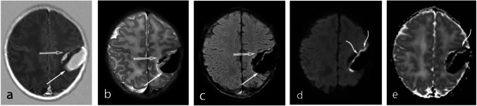
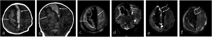
Results: Twenty-eight patients were included in this retrospective study, 82% of whom were born at term. The most common clinical indication for imaging was a combination of apneas and seizures, ocurring in 50%. Hematologic abnormalities were present in 58% of patients. Magnetic resonance imaging (MRI) was performed acutely at the time of presentation between days 1 and 9 of life in 85% of cases. Subpial hemorrhages were unilateral in 86% of neonates, most commonly located in the temporal lobe (44%), and associated with other type of intracranial hemorrhage in 96% of cases, most often parenchymal (86%) and subdural (64%) hemorrhages. We identified three imaging patterns of subpial hemorrhage and two patterns of changes in the underlying brain parenchyma. Additionally, the hyperintense pia mater sign (HPm-sign) was observed on time-of-flight MR angiography (TOF-MRA) in 12 of 18 patients. Neurologic sequelae were noted in 28% of survivors.
Conclusion: Subpial hemorrhage has a distinctive MR pattern, often accompanied with cortical infarction and in most cases underlying parenchymal hemorrhage. In this study, we identified the HPm-sign that may be used to differentiate subpial hemorrhage from other types of hemorrhages. Additionally, we found a correlation between prominent medullary veins (PMV) and intraparenchymal hemorrhage (IPH).
期刊介绍:
Neuroradiology aims to provide state-of-the-art medical and scientific information in the fields of Neuroradiology, Neurosciences, Neurology, Psychiatry, Neurosurgery, and related medical specialities. Neuroradiology as the official Journal of the European Society of Neuroradiology receives submissions from all parts of the world and publishes peer-reviewed original research, comprehensive reviews, educational papers, opinion papers, and short reports on exceptional clinical observations and new technical developments in the field of Neuroimaging and Neurointervention. The journal has subsections for Diagnostic and Interventional Neuroradiology, Advanced Neuroimaging, Paediatric Neuroradiology, Head-Neck-ENT Radiology, Spine Neuroradiology, and for submissions from Japan. Neuroradiology aims to provide new knowledge about and insights into the function and pathology of the human nervous system that may help to better diagnose and treat nervous system diseases. Neuroradiology is a member of the Committee on Publication Ethics (COPE) and follows the COPE core practices. Neuroradiology prefers articles that are free of bias, self-critical regarding limitations, transparent and clear in describing study participants, methods, and statistics, and short in presenting results. Before peer-review all submissions are automatically checked by iThenticate to assess for potential overlap in prior publication.


 分享
分享
 求助内容:
求助内容: 应助结果提醒方式:
 扫码关注我们
