Quinn Steiner, Albert Wang, Laura Slane, Scott Hetzel, Ryan DeWall, Darryl Thelen, Kenneth Lee
{"title":"Ultrasound quantitative characterization of tendinopathy with shear wave elastography in an ex vivo porcine tendon model.","authors":"Quinn Steiner, Albert Wang, Laura Slane, Scott Hetzel, Ryan DeWall, Darryl Thelen, Kenneth Lee","doi":"10.1186/s41747-024-00542-1","DOIUrl":null,"url":null,"abstract":"<p><strong>Background: </strong>Early detection and treatment of tendinopathy may prevent progression to partial tears or complete rupture. Shear wave elastography (SWE) may help address the need for better tendon pathology characterization. This study aimed to quantify the effect of structural damage in an ex vivo animal tendinopathy model using SWE.</p><p><strong>Methods: </strong>Forty-two porcine flexor tendons were injected with a 0.05-mL bolus of 1.5% collagenase solution to induce focal structural damage without surface tears. Control tendons were injected with saline (n = 42). Twenty-one tendons from each group were incubated at 37 °C for 3.5 h, while the remaining 21 from each group were incubated for 7 h. Each group was then divided into three groups of seven, and tendon incisions were made at 25%, 50%, and 75% of the tendon thickness. Tendons were mechanically stretched axially during simultaneous collection of SWE at the injection site.</p><p><strong>Results: </strong>There were significant differences in shear wave speed (SWS) (saline > collagenase) at 3.5-h incubation (p < 0.001) and 7-h incubation (p < 0.001). Additionally, there was a significant difference in SWS between tendons cut at 25% and tendons cut at 50% and 75% (p = 0.040 and p = 0.001, respectively). Collagenase-treated tendons ruptured at a lower force than saline-treated tendons at both incubation times (both p < 0.001) when controlling for cut depth. Tendons treated with collagenase ruptured at a lower force than the saline control group at each cut thickness (all p < 0.001) controlling for incubation time.</p><p><strong>Conclusion: </strong>In a controlled ex vivo porcine model, SWE can be used to detect structural damage associated with tendinopathy.</p><p><strong>Relevance statement: </strong>Shear wave elastography can be used to show differences in abnormal tendons that may be translatable to clinical use as an adjunctive measure of tendon elasticity and injury.</p><p><strong>Key points: </strong>Tendon abnormality was quantitatively characterized using shear wave elastography in an ex vivo porcine experimental model. Shear wave speed was an accurate imaging biomarker for tendon health. Shear wave elastography was effective at detecting the extent of tendon damage. Tendons with decreased shear wave speed measurements rupture at smaller applied mechanical force.</p>","PeriodicalId":36926,"journal":{"name":"European Radiology Experimental","volume":"9 1","pages":"33"},"PeriodicalIF":3.6000,"publicationDate":"2025-03-20","publicationTypes":"Journal Article","fieldsOfStudy":null,"isOpenAccess":false,"openAccessPdf":"https://www.ncbi.nlm.nih.gov/pmc/articles/PMC11926283/pdf/","citationCount":"0","resultStr":null,"platform":"Semanticscholar","paperid":null,"PeriodicalName":"European Radiology Experimental","FirstCategoryId":"1085","ListUrlMain":"https://doi.org/10.1186/s41747-024-00542-1","RegionNum":0,"RegionCategory":null,"ArticlePicture":[],"TitleCN":null,"AbstractTextCN":null,"PMCID":null,"EPubDate":"","PubModel":"","JCR":"Q1","JCRName":"RADIOLOGY, NUCLEAR MEDICINE & MEDICAL IMAGING","Score":null,"Total":0}
引用次数: 0
Abstract
Background: Early detection and treatment of tendinopathy may prevent progression to partial tears or complete rupture. Shear wave elastography (SWE) may help address the need for better tendon pathology characterization. This study aimed to quantify the effect of structural damage in an ex vivo animal tendinopathy model using SWE.
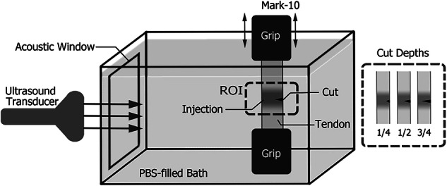
Methods: Forty-two porcine flexor tendons were injected with a 0.05-mL bolus of 1.5% collagenase solution to induce focal structural damage without surface tears. Control tendons were injected with saline (n = 42). Twenty-one tendons from each group were incubated at 37 °C for 3.5 h, while the remaining 21 from each group were incubated for 7 h. Each group was then divided into three groups of seven, and tendon incisions were made at 25%, 50%, and 75% of the tendon thickness. Tendons were mechanically stretched axially during simultaneous collection of SWE at the injection site.
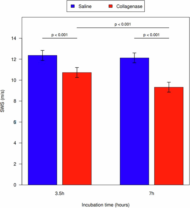
Results: There were significant differences in shear wave speed (SWS) (saline > collagenase) at 3.5-h incubation (p < 0.001) and 7-h incubation (p < 0.001). Additionally, there was a significant difference in SWS between tendons cut at 25% and tendons cut at 50% and 75% (p = 0.040 and p = 0.001, respectively). Collagenase-treated tendons ruptured at a lower force than saline-treated tendons at both incubation times (both p < 0.001) when controlling for cut depth. Tendons treated with collagenase ruptured at a lower force than the saline control group at each cut thickness (all p < 0.001) controlling for incubation time.
Conclusion: In a controlled ex vivo porcine model, SWE can be used to detect structural damage associated with tendinopathy.
Relevance statement: Shear wave elastography can be used to show differences in abnormal tendons that may be translatable to clinical use as an adjunctive measure of tendon elasticity and injury.
Key points: Tendon abnormality was quantitatively characterized using shear wave elastography in an ex vivo porcine experimental model. Shear wave speed was an accurate imaging biomarker for tendon health. Shear wave elastography was effective at detecting the extent of tendon damage. Tendons with decreased shear wave speed measurements rupture at smaller applied mechanical force.


 分享
分享
 求助内容:
求助内容: 应助结果提醒方式:
 扫码关注我们
