Danielle E Whittier, Matthias Walle, Penny R Atkins, Caitlyn J Collins, Matthias A Zumstein, Patrik Christen, Kurt Lippuner, Ralph Müller
{"title":"Structural alterations during fracture healing lead to void spaces developing in surrounding bone microarchitecture.","authors":"Danielle E Whittier, Matthias Walle, Penny R Atkins, Caitlyn J Collins, Matthias A Zumstein, Patrik Christen, Kurt Lippuner, Ralph Müller","doi":"10.1093/jbmr/zjaf046","DOIUrl":null,"url":null,"abstract":"<p><p>Distal radius fractures are among the most common fracture sites, with a high incidence across all age groups. High-resolution peripheral quantitative computed tomography (HR-pQCT) has enabled assessment of bone microarchitecture in vivo at the distal radius, providing new insights into the healing process. However, we have observed structural bone loss that is not captured by standard analysis. This study uses void space analysis to quantify the development of localized structural bone loss during fracture healing. Twenty-six participants (21 female, 5 male; aged 18-79 yr) with conservatively-treated distal radius fractures were scanned using HR-pQCT at 6 study visits post-fracture (wk 1, 3, 5, 12, 26, and 52). Total BMD (Tt.BMD), bone volume fraction (BV/TV), and void space volume fraction (VS/TV) were measured. Grip strength relative to the non-fractured wrist and patient rated wrist evaluation (PRWE) were measured at all study visits after cast removal. The cumulative expansion of VS/TV across sequential study visits was quantified to differentiate voids that developed during healing from pre-existing void space. A 5-fold increase in median VS/TV was observed during the follow-up period, from 1.0% (0.6%-9.0%) to 5.5% (2.5%-12.4%). Tt.BMD and BV/TV did not significantly change in this same time interval. Relative grip strength after cast removal was significantly inversely correlated with final VS/TV (⍴ = -0.63, p = .02) and cumulative expansion of new void space during healing (R = -0.67, p <.01), whereas no significant associations were found with age or PRWE. This study suggests that there are adverse changes in bone microarchitecture during fracture healing, despite the preservation of overall Tt.BMD and BV/TV in the same region. Reduced grip strength is correlated with more severe void space formation, but the mechanistic relationship requires further exploration. The formation of void spaces may have long-term implications on bone strength and could provide insight into risk of re-fracture.</p>","PeriodicalId":185,"journal":{"name":"Journal of Bone and Mineral Research","volume":" ","pages":"791-798"},"PeriodicalIF":5.9000,"publicationDate":"2025-06-03","publicationTypes":"Journal Article","fieldsOfStudy":null,"isOpenAccess":false,"openAccessPdf":"https://www.ncbi.nlm.nih.gov/pmc/articles/PMC12131236/pdf/","citationCount":"0","resultStr":null,"platform":"Semanticscholar","paperid":null,"PeriodicalName":"Journal of Bone and Mineral Research","FirstCategoryId":"3","ListUrlMain":"https://doi.org/10.1093/jbmr/zjaf046","RegionNum":1,"RegionCategory":"医学","ArticlePicture":[],"TitleCN":null,"AbstractTextCN":null,"PMCID":null,"EPubDate":"","PubModel":"","JCR":"Q1","JCRName":"ENDOCRINOLOGY & METABOLISM","Score":null,"Total":0}
引用次数: 0
Abstract
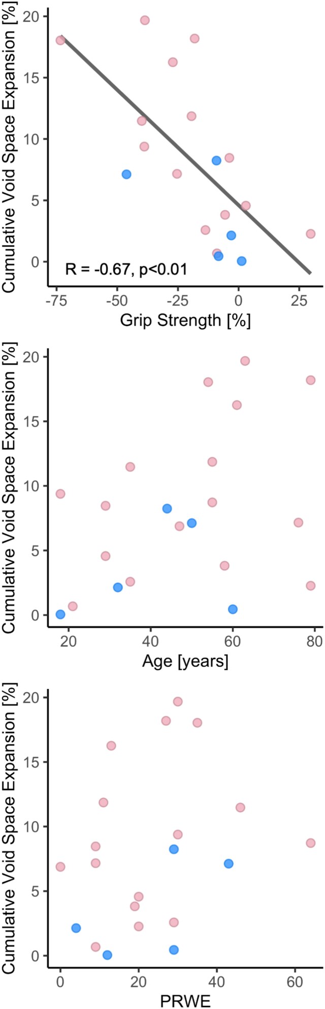
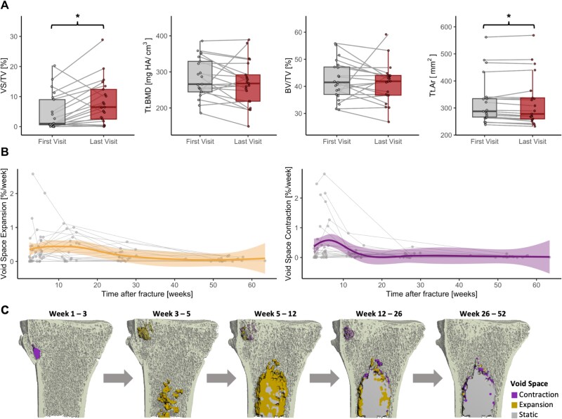
Distal radius fractures are among the most common fracture sites, with a high incidence across all age groups. High-resolution peripheral quantitative computed tomography (HR-pQCT) has enabled assessment of bone microarchitecture in vivo at the distal radius, providing new insights into the healing process. However, we have observed structural bone loss that is not captured by standard analysis. This study uses void space analysis to quantify the development of localized structural bone loss during fracture healing. Twenty-six participants (21 female, 5 male; aged 18-79 yr) with conservatively-treated distal radius fractures were scanned using HR-pQCT at 6 study visits post-fracture (wk 1, 3, 5, 12, 26, and 52). Total BMD (Tt.BMD), bone volume fraction (BV/TV), and void space volume fraction (VS/TV) were measured. Grip strength relative to the non-fractured wrist and patient rated wrist evaluation (PRWE) were measured at all study visits after cast removal. The cumulative expansion of VS/TV across sequential study visits was quantified to differentiate voids that developed during healing from pre-existing void space. A 5-fold increase in median VS/TV was observed during the follow-up period, from 1.0% (0.6%-9.0%) to 5.5% (2.5%-12.4%). Tt.BMD and BV/TV did not significantly change in this same time interval. Relative grip strength after cast removal was significantly inversely correlated with final VS/TV (⍴ = -0.63, p = .02) and cumulative expansion of new void space during healing (R = -0.67, p <.01), whereas no significant associations were found with age or PRWE. This study suggests that there are adverse changes in bone microarchitecture during fracture healing, despite the preservation of overall Tt.BMD and BV/TV in the same region. Reduced grip strength is correlated with more severe void space formation, but the mechanistic relationship requires further exploration. The formation of void spaces may have long-term implications on bone strength and could provide insight into risk of re-fracture.
期刊介绍:
The Journal of Bone and Mineral Research (JBMR) publishes highly impactful original manuscripts, reviews, and special articles on basic, translational and clinical investigations relevant to the musculoskeletal system and mineral metabolism. Specifically, the journal is interested in original research on the biology and physiology of skeletal tissues, interdisciplinary research spanning the musculoskeletal and other systems, including but not limited to immunology, hematology, energy metabolism, cancer biology, and neurology, and systems biology topics using large scale “-omics” approaches. The journal welcomes clinical research on the pathophysiology, treatment and prevention of osteoporosis and fractures, as well as sarcopenia, disorders of bone and mineral metabolism, and rare or genetically determined bone diseases.


 分享
分享
 求助内容:
求助内容: 应助结果提醒方式:
 扫码关注我们
