Destie Provenzano, Jeffrey Wang, Sharad Goyal, Yuan James Rao
{"title":"Discussion of a Simple Method to Generate Descriptive Images Using Predictive ResNet Model Weights and Feature Maps for Recurrent Cervix Cancer.","authors":"Destie Provenzano, Jeffrey Wang, Sharad Goyal, Yuan James Rao","doi":"10.3390/tomography11030038","DOIUrl":null,"url":null,"abstract":"<p><strong>Background: </strong>Predictive models like Residual Neural Networks (ResNets) can use Magnetic Resonance Imaging (MRI) data to identify cervix tumors likely to recur after radiotherapy (RT) with high accuracy. However, there persists a lack of insight into model selections (explainability). In this study, we explored whether model features could be used to generate simulated images as a method of model explainability.</p><p><strong>Methods: </strong>T2W MRI data were collected for twenty-seven women with cervix cancer who received RT from the TCGA-CESC database. Simulated images were generated as follows: [A] a ResNet model was trained to identify recurrent cervix cancer; [B] a model was evaluated on T2W MRI data for subjects to obtain corresponding feature maps; [C] most important feature maps were determined for each image; [D] feature maps were combined across all images to generate a simulated image; [E] the final image was reviewed by a radiation oncologist and an initial algorithm to identify the likelihood of recurrence.</p><p><strong>Results: </strong>Predictive feature maps from the ResNet model (93% accuracy) were used to generate simulated images. Simulated images passed through the model were identified as recurrent and non-recurrent cervix tumors after radiotherapy. A radiation oncologist identified the simulated images as cervix tumors with characteristics of aggressive Cervical Cancer. These images also contained multiple MRI features not considered clinically relevant.</p><p><strong>Conclusion: </strong>This simple method was able to generate simulated MRI data that mimicked recurrent and non-recurrent cervix cancer tumor images. These generated images could be useful for evaluating the explainability of predictive models and to assist radiologists with the identification of features likely to predict disease course.</p>","PeriodicalId":51330,"journal":{"name":"Tomography","volume":"11 3","pages":""},"PeriodicalIF":2.2000,"publicationDate":"2025-03-20","publicationTypes":"Journal Article","fieldsOfStudy":null,"isOpenAccess":false,"openAccessPdf":"https://www.ncbi.nlm.nih.gov/pmc/articles/PMC11946054/pdf/","citationCount":"0","resultStr":null,"platform":"Semanticscholar","paperid":null,"PeriodicalName":"Tomography","FirstCategoryId":"3","ListUrlMain":"https://doi.org/10.3390/tomography11030038","RegionNum":4,"RegionCategory":"医学","ArticlePicture":[],"TitleCN":null,"AbstractTextCN":null,"PMCID":null,"EPubDate":"","PubModel":"","JCR":"Q2","JCRName":"RADIOLOGY, NUCLEAR MEDICINE & MEDICAL IMAGING","Score":null,"Total":0}
引用次数: 0
Abstract
Background: Predictive models like Residual Neural Networks (ResNets) can use Magnetic Resonance Imaging (MRI) data to identify cervix tumors likely to recur after radiotherapy (RT) with high accuracy. However, there persists a lack of insight into model selections (explainability). In this study, we explored whether model features could be used to generate simulated images as a method of model explainability.
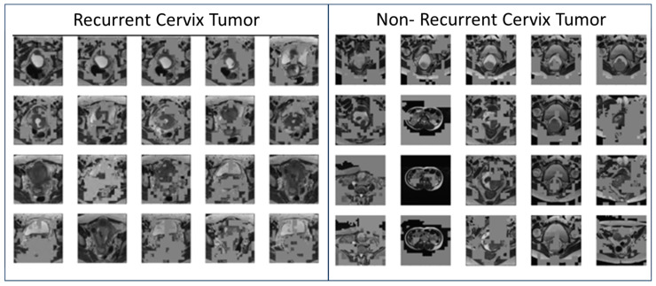
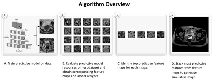
Methods: T2W MRI data were collected for twenty-seven women with cervix cancer who received RT from the TCGA-CESC database. Simulated images were generated as follows: [A] a ResNet model was trained to identify recurrent cervix cancer; [B] a model was evaluated on T2W MRI data for subjects to obtain corresponding feature maps; [C] most important feature maps were determined for each image; [D] feature maps were combined across all images to generate a simulated image; [E] the final image was reviewed by a radiation oncologist and an initial algorithm to identify the likelihood of recurrence.
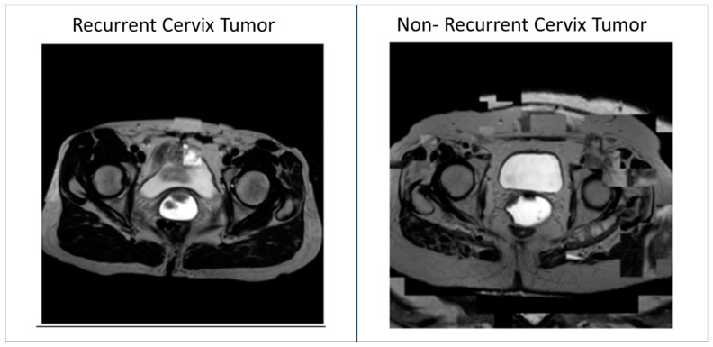
Results: Predictive feature maps from the ResNet model (93% accuracy) were used to generate simulated images. Simulated images passed through the model were identified as recurrent and non-recurrent cervix tumors after radiotherapy. A radiation oncologist identified the simulated images as cervix tumors with characteristics of aggressive Cervical Cancer. These images also contained multiple MRI features not considered clinically relevant.
Conclusion: This simple method was able to generate simulated MRI data that mimicked recurrent and non-recurrent cervix cancer tumor images. These generated images could be useful for evaluating the explainability of predictive models and to assist radiologists with the identification of features likely to predict disease course.
TomographyMedicine-Radiology, Nuclear Medicine and Imaging
CiteScore
2.70
自引率
10.50%
发文量
222
期刊介绍:
TomographyTM publishes basic (technical and pre-clinical) and clinical scientific articles which involve the advancement of imaging technologies. Tomography encompasses studies that use single or multiple imaging modalities including for example CT, US, PET, SPECT, MR and hyperpolarization technologies, as well as optical modalities (i.e. bioluminescence, photoacoustic, endomicroscopy, fiber optic imaging and optical computed tomography) in basic sciences, engineering, preclinical and clinical medicine.
Tomography also welcomes studies involving exploration and refinement of contrast mechanisms and image-derived metrics within and across modalities toward the development of novel imaging probes for image-based feedback and intervention. The use of imaging in biology and medicine provides unparalleled opportunities to noninvasively interrogate tissues to obtain real-time dynamic and quantitative information required for diagnosis and response to interventions and to follow evolving pathological conditions. As multi-modal studies and the complexities of imaging technologies themselves are ever increasing to provide advanced information to scientists and clinicians.
Tomography provides a unique publication venue allowing investigators the opportunity to more precisely communicate integrated findings related to the diverse and heterogeneous features associated with underlying anatomical, physiological, functional, metabolic and molecular genetic activities of normal and diseased tissue. Thus Tomography publishes peer-reviewed articles which involve the broad use of imaging of any tissue and disease type including both preclinical and clinical investigations. In addition, hardware/software along with chemical and molecular probe advances are welcome as they are deemed to significantly contribute towards the long-term goal of improving the overall impact of imaging on scientific and clinical discovery.


 分享
分享
 求助内容:
求助内容: 应助结果提醒方式:
 扫码关注我们
