Neurological disability and brain grey matter atrophy in primary progressive multiple sclerosis are determined by microstructural lesional changes, but not by lesion load.
Theodoros Ladopoulos, Zainab Abbas, Britta Krieger, Barbara Bellenberg, Jeyanthan Charles James, Jana Bauer, Ralf Gold, Carsten Lukas, Ruth Schneider
{"title":"Neurological disability and brain grey matter atrophy in primary progressive multiple sclerosis are determined by microstructural lesional changes, but not by lesion load.","authors":"Theodoros Ladopoulos, Zainab Abbas, Britta Krieger, Barbara Bellenberg, Jeyanthan Charles James, Jana Bauer, Ralf Gold, Carsten Lukas, Ruth Schneider","doi":"10.1007/s00415-025-13043-x","DOIUrl":null,"url":null,"abstract":"<p><strong>Background: </strong>Conventional MRI measures, such as the number and volume of MS lesions, are histologically non-specific and cannot sufficiently explain clinical disability or brain atrophy in MS. Nevertheless, demyelinating plaques exhibit distinct histopathological features in relapsing and progressive multiple sclerosis (MS) subtypes. The aim of this study was to assess microstructural characteristics of MS lesions using quantitative MRI and explore their associations with grey matter (GM) atrophy and clinical disability.</p><p><strong>Methods: </strong>56 control subjects (CS), 121 patients with relapsing-remitting (RRMS), and 38 patients with primary progressive MS (PPMS) underwent 1.5 T MRI scans and clinical examinations. Lesion and brain segmentation based on T1-weighted and FLAIR images were performed using SAMSEG. The MDME sequence and SyMRI software were used to estimate relaxation rates and myelin volume fraction in MS lesions and normal-appearing white matter (NAWM). Associations between quantitative lesional and NAWM MRI parameters with GM atrophy and clinical disability were investigated.</p><p><strong>Results: </strong>Brain regional volumes and quantitative lesional and NAWM MRI parameters were significantly decreased in patients with PPMS compared to those with RRMS. Quantitative lesional MRI parameters demonstrated statistically significant associations with cortical and deep GM volumes as well as with disability scores in RRMS and especially in PPMS. In contrast to RRMS, lesion volume was not associated with either GM atrophy or clinical disability in the PPMS group.</p><p><strong>Conclusions: </strong>Quantitative lesional MRI measures, but not lesion load, were strongly associated with clinical disability and GM atrophy in PPMS patients, likely reflecting differences in lesion pathology between MS subtypes.</p>","PeriodicalId":16558,"journal":{"name":"Journal of Neurology","volume":"272 4","pages":"302"},"PeriodicalIF":4.6000,"publicationDate":"2025-04-01","publicationTypes":"Journal Article","fieldsOfStudy":null,"isOpenAccess":false,"openAccessPdf":"https://www.ncbi.nlm.nih.gov/pmc/articles/PMC11961454/pdf/","citationCount":"0","resultStr":null,"platform":"Semanticscholar","paperid":null,"PeriodicalName":"Journal of Neurology","FirstCategoryId":"3","ListUrlMain":"https://doi.org/10.1007/s00415-025-13043-x","RegionNum":2,"RegionCategory":"医学","ArticlePicture":[],"TitleCN":null,"AbstractTextCN":null,"PMCID":null,"EPubDate":"","PubModel":"","JCR":"Q1","JCRName":"CLINICAL NEUROLOGY","Score":null,"Total":0}
引用次数: 0
Abstract
Background: Conventional MRI measures, such as the number and volume of MS lesions, are histologically non-specific and cannot sufficiently explain clinical disability or brain atrophy in MS. Nevertheless, demyelinating plaques exhibit distinct histopathological features in relapsing and progressive multiple sclerosis (MS) subtypes. The aim of this study was to assess microstructural characteristics of MS lesions using quantitative MRI and explore their associations with grey matter (GM) atrophy and clinical disability.
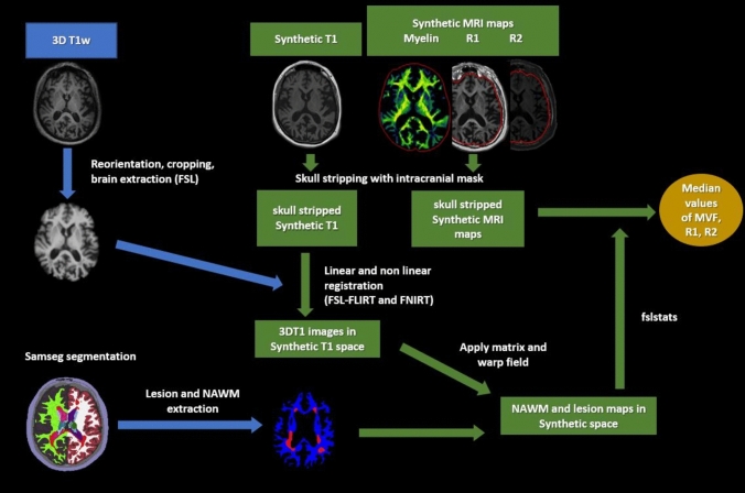
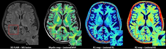
Methods: 56 control subjects (CS), 121 patients with relapsing-remitting (RRMS), and 38 patients with primary progressive MS (PPMS) underwent 1.5 T MRI scans and clinical examinations. Lesion and brain segmentation based on T1-weighted and FLAIR images were performed using SAMSEG. The MDME sequence and SyMRI software were used to estimate relaxation rates and myelin volume fraction in MS lesions and normal-appearing white matter (NAWM). Associations between quantitative lesional and NAWM MRI parameters with GM atrophy and clinical disability were investigated.
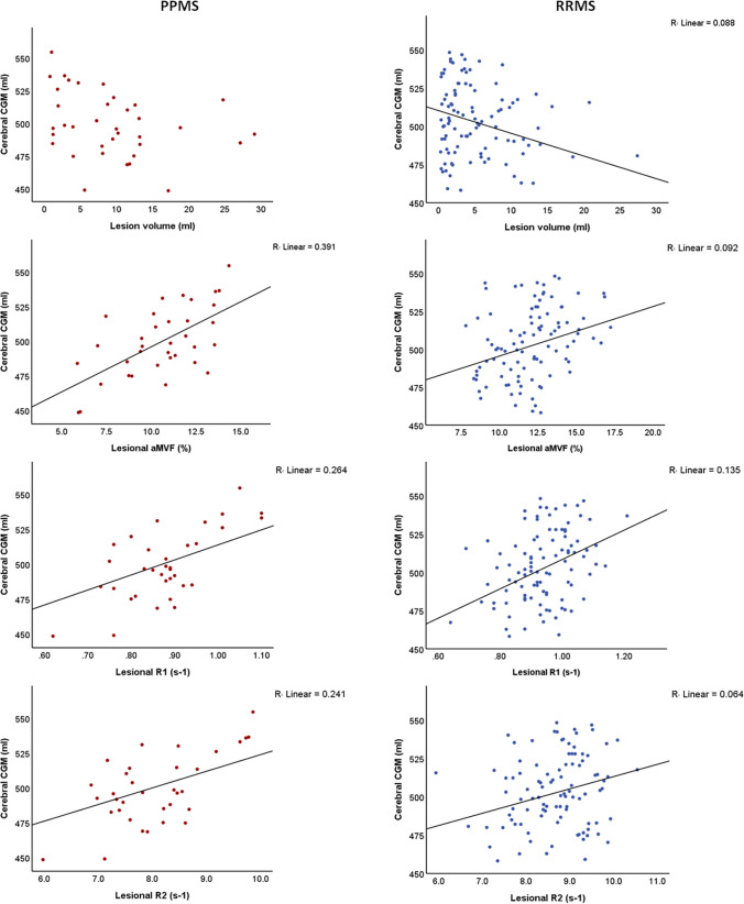
Results: Brain regional volumes and quantitative lesional and NAWM MRI parameters were significantly decreased in patients with PPMS compared to those with RRMS. Quantitative lesional MRI parameters demonstrated statistically significant associations with cortical and deep GM volumes as well as with disability scores in RRMS and especially in PPMS. In contrast to RRMS, lesion volume was not associated with either GM atrophy or clinical disability in the PPMS group.
Conclusions: Quantitative lesional MRI measures, but not lesion load, were strongly associated with clinical disability and GM atrophy in PPMS patients, likely reflecting differences in lesion pathology between MS subtypes.
期刊介绍:
The Journal of Neurology is an international peer-reviewed journal which provides a source for publishing original communications and reviews on clinical neurology covering the whole field.
In addition, Letters to the Editors serve as a forum for clinical cases and the exchange of ideas which highlight important new findings. A section on Neurological progress serves to summarise the major findings in certain fields of neurology. Commentaries on new developments in clinical neuroscience, which may be commissioned or submitted, are published as editorials.
Every neurologist interested in the current diagnosis and treatment of neurological disorders needs access to the information contained in this valuable journal.


 分享
分享
 求助内容:
求助内容: 应助结果提醒方式:
 扫码关注我们
