Zhaohui Jin, Lei Tian, Yangyang Li, Dong Wang, Lei Tang, Ran Wang, Feiyu Ding, Chengyuan Huang, Kun Yang
{"title":"FET-CREB fusion-positive extra-axial myxoid mesenchymal tumor in the cerebellum: illustrative case.","authors":"Zhaohui Jin, Lei Tian, Yangyang Li, Dong Wang, Lei Tang, Ran Wang, Feiyu Ding, Chengyuan Huang, Kun Yang","doi":"10.3171/CASE24872","DOIUrl":null,"url":null,"abstract":"<p><strong>Background: </strong>Myxoid mesenchymal tumor (MMT) is an exceptionally rare central nervous system (CNS) tumor, with even fewer reported cases in the cerebellum. Its complex histopathological features and nonspecific clinical presentation pose considerable challenges in diagnosis. The rarity of the tumor, coupled with its poorly characterized clinical and radiological features, complicates early detection and effective treatment.</p><p><strong>Observations: </strong>xsThe authors present the case of an 18-year-old female who presented with persistent headaches and intermittent diplopia. MRI revealed a hypervascular mass in the right cerebellum, showing marked contrast enhancement. The patient underwent total tumor resection, and histopathological examination revealed lobulated tumor cells that were positive for the FET-CREB fusion gene. Immunohistochemical staining was positive for epithelial membrane antigen, vimentin, and H3K27me3, with a Ki-67 proliferation index of 8%, confirming the diagnosis of MMT. The patient had an uneventful recovery and remained recurrence free during a 6-month follow-up.</p><p><strong>Lessons: </strong>This case highlights the critical role of the FET-CREB fusion gene in diagnosing cerebellar MMT. It emphasizes the importance of early recognition, comprehensive pathological evaluation, and genetic analysis in managing this rare tumor. A thorough, multidisciplinary diagnostic approach is essential for determining the optimal treatment and improving patient outcomes. https://thejns.org/doi/10.3171/CASE24872.</p>","PeriodicalId":94098,"journal":{"name":"Journal of neurosurgery. Case lessons","volume":"9 13","pages":""},"PeriodicalIF":0.0000,"publicationDate":"2025-03-31","publicationTypes":"Journal Article","fieldsOfStudy":null,"isOpenAccess":false,"openAccessPdf":"https://www.ncbi.nlm.nih.gov/pmc/articles/PMC11959636/pdf/","citationCount":"0","resultStr":null,"platform":"Semanticscholar","paperid":null,"PeriodicalName":"Journal of neurosurgery. Case lessons","FirstCategoryId":"1085","ListUrlMain":"https://doi.org/10.3171/CASE24872","RegionNum":0,"RegionCategory":null,"ArticlePicture":[],"TitleCN":null,"AbstractTextCN":null,"PMCID":null,"EPubDate":"","PubModel":"","JCR":"","JCRName":"","Score":null,"Total":0}
引用次数: 0
Abstract
Background: Myxoid mesenchymal tumor (MMT) is an exceptionally rare central nervous system (CNS) tumor, with even fewer reported cases in the cerebellum. Its complex histopathological features and nonspecific clinical presentation pose considerable challenges in diagnosis. The rarity of the tumor, coupled with its poorly characterized clinical and radiological features, complicates early detection and effective treatment.
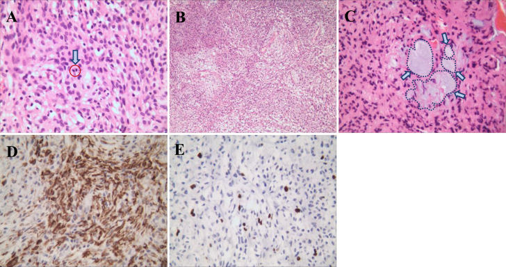
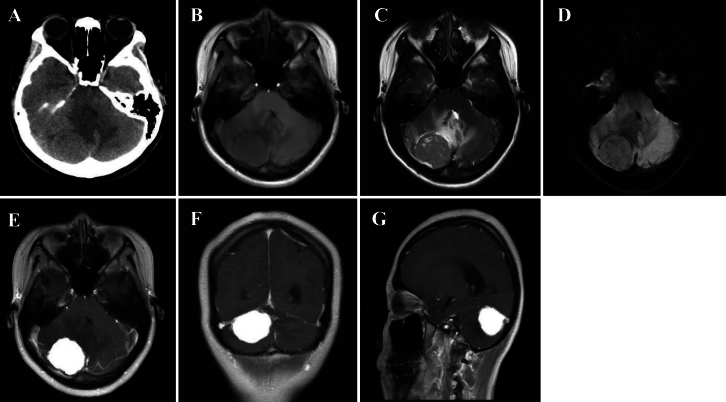
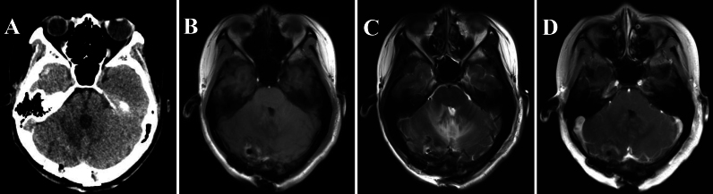
Observations: xsThe authors present the case of an 18-year-old female who presented with persistent headaches and intermittent diplopia. MRI revealed a hypervascular mass in the right cerebellum, showing marked contrast enhancement. The patient underwent total tumor resection, and histopathological examination revealed lobulated tumor cells that were positive for the FET-CREB fusion gene. Immunohistochemical staining was positive for epithelial membrane antigen, vimentin, and H3K27me3, with a Ki-67 proliferation index of 8%, confirming the diagnosis of MMT. The patient had an uneventful recovery and remained recurrence free during a 6-month follow-up.
Lessons: This case highlights the critical role of the FET-CREB fusion gene in diagnosing cerebellar MMT. It emphasizes the importance of early recognition, comprehensive pathological evaluation, and genetic analysis in managing this rare tumor. A thorough, multidisciplinary diagnostic approach is essential for determining the optimal treatment and improving patient outcomes. https://thejns.org/doi/10.3171/CASE24872.


 分享
分享

 求助内容:
求助内容: 应助结果提醒方式:
 扫码关注我们
