Ali Tavoosian, Amirreza Shamshirgaran, Seyed Mohammad Kazem Aghamir
{"title":"Bilateral Renal Infarction, a Rare Consequence of Blunt Renal Artery Injury: A Case Report.","authors":"Ali Tavoosian, Amirreza Shamshirgaran, Seyed Mohammad Kazem Aghamir","doi":"10.1177/11795476241297632","DOIUrl":null,"url":null,"abstract":"<p><strong>Background: </strong>Renal infarction is an uncommon complication of Blunt renal artery injury (BRAI) following abdominal trauma. Diagnosis of infarction is difficult and mostly delayed due to non-specific symptoms. Early diagnosis can lead to appropriate and effective treatment, which prevents further complication.</p><p><strong>Case presentation: </strong>We report a case of 21-year-old man falling from a height of 9 m. A contrast-enhanced CT scan in the nephrogram phase showed no evidence of absorption in the right kidney and significantly decreased absorption in the left kidney. The pyelogram phase showed no secretion in the right kidney and decreased secretion in the left kidney suggesting segmental renal infarction. Subsequently, heparin infusion was initiated immediately. A follow-up contrast- enhanced abdominopelvic CT scan was performed after 1 month and showed no sign of infarction, and all laboratory tests were normal.</p><p><strong>Conclusion: </strong>Contrast-enhanced abdominopelvic CT scan helps physicians diagnose the renal infarction immediately and start appropriate treatment. Treatment can vary from aggressive surgical procedures to observation and supportive care.</p>","PeriodicalId":10357,"journal":{"name":"Clinical Medicine Insights. Case Reports","volume":"18 ","pages":"11795476241297632"},"PeriodicalIF":0.6000,"publicationDate":"2025-03-31","publicationTypes":"Journal Article","fieldsOfStudy":null,"isOpenAccess":false,"openAccessPdf":"https://www.ncbi.nlm.nih.gov/pmc/articles/PMC11960154/pdf/","citationCount":"0","resultStr":null,"platform":"Semanticscholar","paperid":null,"PeriodicalName":"Clinical Medicine Insights. Case Reports","FirstCategoryId":"1085","ListUrlMain":"https://doi.org/10.1177/11795476241297632","RegionNum":0,"RegionCategory":null,"ArticlePicture":[],"TitleCN":null,"AbstractTextCN":null,"PMCID":null,"EPubDate":"2025/1/1 0:00:00","PubModel":"eCollection","JCR":"Q3","JCRName":"MEDICINE, GENERAL & INTERNAL","Score":null,"Total":0}
引用次数: 0
Abstract
Background: Renal infarction is an uncommon complication of Blunt renal artery injury (BRAI) following abdominal trauma. Diagnosis of infarction is difficult and mostly delayed due to non-specific symptoms. Early diagnosis can lead to appropriate and effective treatment, which prevents further complication.
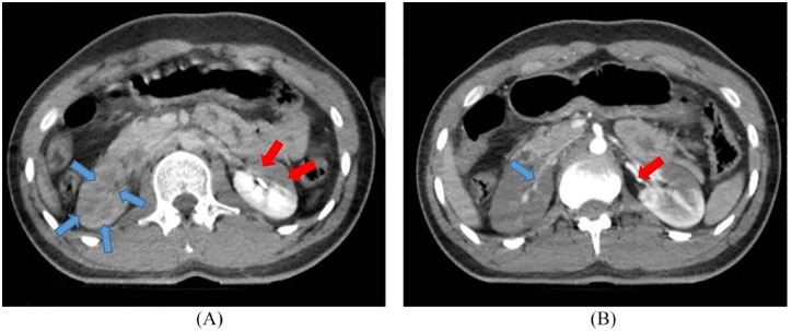
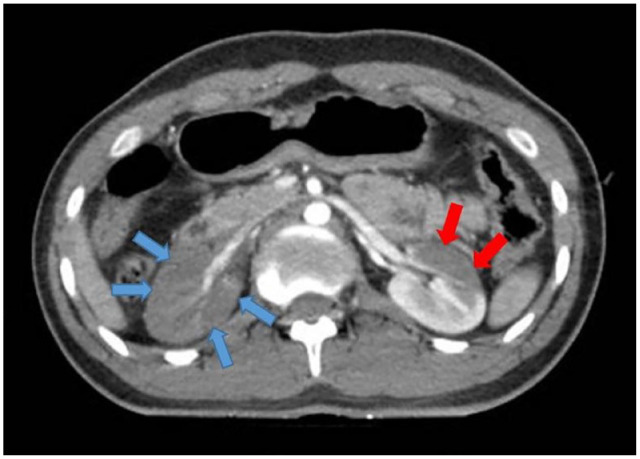
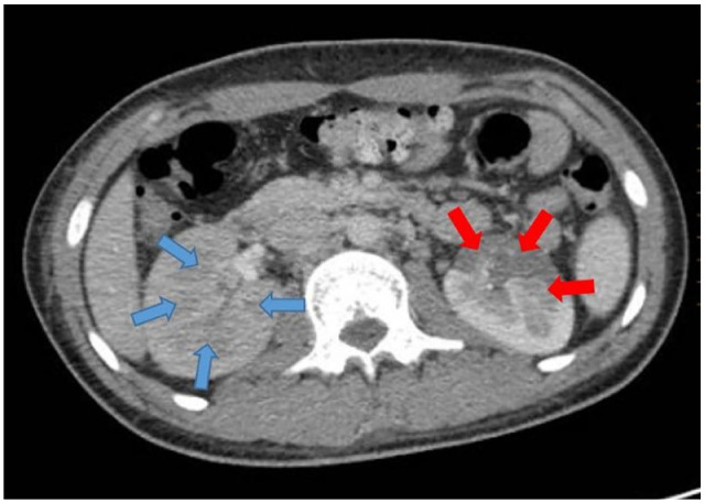
Case presentation: We report a case of 21-year-old man falling from a height of 9 m. A contrast-enhanced CT scan in the nephrogram phase showed no evidence of absorption in the right kidney and significantly decreased absorption in the left kidney. The pyelogram phase showed no secretion in the right kidney and decreased secretion in the left kidney suggesting segmental renal infarction. Subsequently, heparin infusion was initiated immediately. A follow-up contrast- enhanced abdominopelvic CT scan was performed after 1 month and showed no sign of infarction, and all laboratory tests were normal.
Conclusion: Contrast-enhanced abdominopelvic CT scan helps physicians diagnose the renal infarction immediately and start appropriate treatment. Treatment can vary from aggressive surgical procedures to observation and supportive care.


 分享
分享
 求助内容:
求助内容: 应助结果提醒方式:
 扫码关注我们
