Jesper Swärd, Karl Bohlin, Olof Henrikson, Sven Lundstam, Ralph Peeker, Anna Grenabo Bergdahl
{"title":"Renal angiomyolipoma-investigating radiological signs indicative of risk for bleeding.","authors":"Jesper Swärd, Karl Bohlin, Olof Henrikson, Sven Lundstam, Ralph Peeker, Anna Grenabo Bergdahl","doi":"10.1186/s13244-025-01957-z","DOIUrl":null,"url":null,"abstract":"<p><strong>Objectives: </strong>To compare imaging differences between bleeding and non-bleeding angiomyolipoma with respect to the proportion and attenuation of the angiomyogenic component and the occurrence and size of aneurysms.</p><p><strong>Materials and methods: </strong>CT scans and angiographies preceding 58 consecutive embolisations at two institutions from 1999 to 2018 were analysed retrospectively. Tumour volume was measured by contouring the angiomyolipoma on CT scans. The partial volume of the angiomyogenic component (blood vessels and smooth muscle relative to fatty tissue) was derived using attenuation threshold values measured in Hounsfield Units.</p><p><strong>Results: </strong>Bleeding angiomyolipoma exhibited a significantly higher proportion of angiomyogenic component (23%) than non-bleeding angiomyolipoma (8%) (p = 0.042). Angiomyolipoma with 0-5% angiomyogenic component had a lower risk of bleeding compared to those with ≥ 5% angiomyogenic component (13% vs 42%). Mean attenuation values of angiomyogenic components did not differ between bleeders and non-bleeders. Aneurysms were observed in 24% of angiomyolipoma during angiography. No statistically significant association was found between the occurrence of aneurysms and bleeding, neither when all aneurysms were included nor when only aneurysms ≥ 5 mm were considered. Tuberous sclerosis patients had larger tumours (11.4 cm vs 6.0 cm), but no significant difference in bleeding was observed (p = 0.53).</p><p><strong>Conclusions: </strong>A higher proportion of the angiomyogenic component in bleeding renal angiomyolipoma suggests a possible association with bleeding. Angiomyolipoma with less than 5% angiomyogenic components may represent a subgroup with a reduced risk of bleeding. Our findings do not confirm the widely accepted assumption that aneurysms significantly increase the risk of bleeding.</p><p><strong>Critical relevance statement: </strong>Measuring the angiomyogenic component in renal angiomyolipoma could help address current knowledge gaps and aid in the more efficient selection of patients for therapeutic interventions.</p><p><strong>Key points: </strong>Identifying risk factors for bleeding beyond tumour size is important. Very low angiomyogenic component tumours may have reduced bleeding risk. The presence of aneurysms may not significantly increase bleeding risk. Reporting angiomyogenic proportion on CT may aid in treatment decisions.</p>","PeriodicalId":13639,"journal":{"name":"Insights into Imaging","volume":"16 1","pages":"83"},"PeriodicalIF":4.5000,"publicationDate":"2025-04-05","publicationTypes":"Journal Article","fieldsOfStudy":null,"isOpenAccess":false,"openAccessPdf":"https://www.ncbi.nlm.nih.gov/pmc/articles/PMC11972250/pdf/","citationCount":"0","resultStr":null,"platform":"Semanticscholar","paperid":null,"PeriodicalName":"Insights into Imaging","FirstCategoryId":"3","ListUrlMain":"https://doi.org/10.1186/s13244-025-01957-z","RegionNum":2,"RegionCategory":"医学","ArticlePicture":[],"TitleCN":null,"AbstractTextCN":null,"PMCID":null,"EPubDate":"","PubModel":"","JCR":"Q1","JCRName":"RADIOLOGY, NUCLEAR MEDICINE & MEDICAL IMAGING","Score":null,"Total":0}
引用次数: 0
Abstract
Objectives: To compare imaging differences between bleeding and non-bleeding angiomyolipoma with respect to the proportion and attenuation of the angiomyogenic component and the occurrence and size of aneurysms.
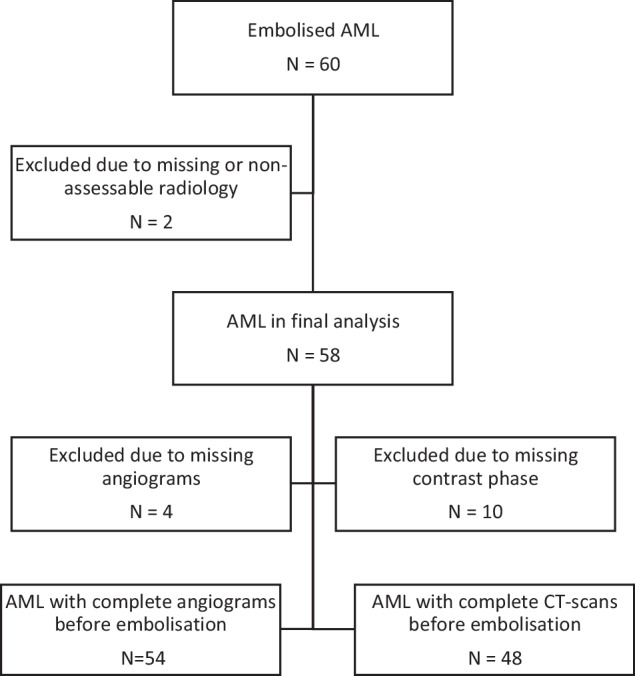
Materials and methods: CT scans and angiographies preceding 58 consecutive embolisations at two institutions from 1999 to 2018 were analysed retrospectively. Tumour volume was measured by contouring the angiomyolipoma on CT scans. The partial volume of the angiomyogenic component (blood vessels and smooth muscle relative to fatty tissue) was derived using attenuation threshold values measured in Hounsfield Units.
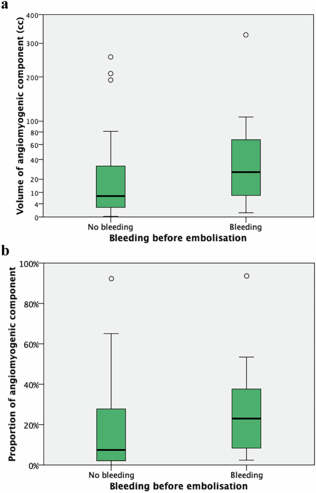
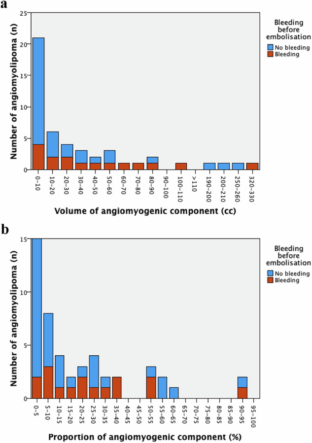
Results: Bleeding angiomyolipoma exhibited a significantly higher proportion of angiomyogenic component (23%) than non-bleeding angiomyolipoma (8%) (p = 0.042). Angiomyolipoma with 0-5% angiomyogenic component had a lower risk of bleeding compared to those with ≥ 5% angiomyogenic component (13% vs 42%). Mean attenuation values of angiomyogenic components did not differ between bleeders and non-bleeders. Aneurysms were observed in 24% of angiomyolipoma during angiography. No statistically significant association was found between the occurrence of aneurysms and bleeding, neither when all aneurysms were included nor when only aneurysms ≥ 5 mm were considered. Tuberous sclerosis patients had larger tumours (11.4 cm vs 6.0 cm), but no significant difference in bleeding was observed (p = 0.53).
Conclusions: A higher proportion of the angiomyogenic component in bleeding renal angiomyolipoma suggests a possible association with bleeding. Angiomyolipoma with less than 5% angiomyogenic components may represent a subgroup with a reduced risk of bleeding. Our findings do not confirm the widely accepted assumption that aneurysms significantly increase the risk of bleeding.
Critical relevance statement: Measuring the angiomyogenic component in renal angiomyolipoma could help address current knowledge gaps and aid in the more efficient selection of patients for therapeutic interventions.
Key points: Identifying risk factors for bleeding beyond tumour size is important. Very low angiomyogenic component tumours may have reduced bleeding risk. The presence of aneurysms may not significantly increase bleeding risk. Reporting angiomyogenic proportion on CT may aid in treatment decisions.
目的:比较出血血管平滑肌脂肪瘤与非出血血管平滑肌脂肪瘤在血管生成成分的比例和衰减、动脉瘤的发生和大小等影像学上的差异。材料和方法:回顾性分析1999年至2018年两家机构连续58例栓塞前的CT扫描和血管造影。通过CT扫描血管平滑肌脂肪瘤的轮廓来测量肿瘤体积。血管生成成分(相对于脂肪组织的血管和平滑肌)的部分体积是通过在霍斯菲尔德单位测量的衰减阈值得出的。结果:出血性血管平滑肌脂肪瘤中血管生成成分的比例(23%)明显高于非出血性血管平滑肌脂肪瘤(8%)(p = 0.042)。与血管生成成分≥5%的血管平滑肌脂肪瘤相比,0-5%血管生成成分的血管平滑肌脂肪瘤出血风险较低(13% vs 42%)。血管生成成分的平均衰减值在出血患者和非出血患者之间没有差异。24%的血管平滑肌脂肪瘤在血管造影中发现动脉瘤。无论是包括所有动脉瘤还是仅考虑≥5mm的动脉瘤,均未发现动脉瘤发生与出血之间有统计学意义的关联。结节性硬化症患者肿瘤较大(11.4 cm vs 6.0 cm),但出血无显著差异(p = 0.53)。结论:出血性肾血管平滑肌脂肪瘤中血管生成成分的比例较高,表明可能与出血有关。血管平滑肌脂肪瘤少于5%的血管生成成分可能代表出血风险降低的亚组。我们的发现并没有证实人们普遍接受的假设,即动脉瘤会显著增加出血的风险。关键相关性声明:测量肾血管平滑肌脂肪瘤的血管生成成分有助于解决目前的知识空白,并有助于更有效地选择患者进行治疗干预。重点:确定肿瘤大小以外出血的危险因素是很重要的。非常低的血管生成成分肿瘤可能降低出血风险。动脉瘤的存在可能不会显著增加出血的风险。在CT上报告血管生成比例可能有助于治疗决策。
期刊介绍:
Insights into Imaging (I³) is a peer-reviewed open access journal published under the brand SpringerOpen. All content published in the journal is freely available online to anyone, anywhere!
I³ continuously updates scientific knowledge and progress in best-practice standards in radiology through the publication of original articles and state-of-the-art reviews and opinions, along with recommendations and statements from the leading radiological societies in Europe.
Founded by the European Society of Radiology (ESR), I³ creates a platform for educational material, guidelines and recommendations, and a forum for topics of controversy.
A balanced combination of review articles, original papers, short communications from European radiological congresses and information on society matters makes I³ an indispensable source for current information in this field.
I³ is owned by the ESR, however authors retain copyright to their article according to the Creative Commons Attribution License (see Copyright and License Agreement). All articles can be read, redistributed and reused for free, as long as the author of the original work is cited properly.
The open access fees (article-processing charges) for this journal are kindly sponsored by ESR for all Members.
The journal went open access in 2012, which means that all articles published since then are freely available online.


 分享
分享
 求助内容:
求助内容: 应助结果提醒方式:
 扫码关注我们
