Pavel Pavlov, Andreas Kontny, Neele Wagner, Nikola Kolev, Alexander Zlatarov, Turgay Kalinov, Anton B Tonchev
{"title":"3D visualization of human colon tissue using a modified CUBIC-based tissue-clearing technique.","authors":"Pavel Pavlov, Andreas Kontny, Neele Wagner, Nikola Kolev, Alexander Zlatarov, Turgay Kalinov, Anton B Tonchev","doi":"10.14440/jbm.2025.0101","DOIUrl":null,"url":null,"abstract":"<p><strong>Background: </strong>Colorectal cancer represents one of the most common neoplastic diseases worldwide, making it a frequent focus in routine pathological analyses. Visualizing complex three-dimensional (3D) structures, such as nerves within tumors, requires thick tissue sections, which necessitates the use of optical tissue-clearing methods to achieve transparency. However, following tissue clearing, samples typically require advanced imaging techniques such as light-sheet and two-photon confocal microscopy, which are usually unavailable in standard histological laboratories.</p><p><strong>Objective: </strong>We aimed to demonstrate how a well-established tissue-clearing approach can be adapted for use in a routine histological laboratory, enabling a robust 3D visualization of nerve fibers in samples of both normal human colon and colon cancer tissues.</p><p><strong>Methods: </strong>We modified the \"clear unobstructed brain/body imaging cocktails\" method, originally developed for whole-brain imaging in mice, and applied it to human colon tissue samples measuring approximately 10 mm<sup>3</sup>, a standard size typically processed in pathological laboratories.</p><p><strong>Results: </strong>Our protocol, which integrates a tissue-clearing technique, enabled reliable immunofluorescent visualization of colonic nerve fibers labeled with anti-β<sub>3</sub>-tubulin antibodies. The labeled nerve fibers could be observed using a standard epifluorescence microscope, and high-quality 3D reconstructions were generated through a simple image analysis approach using the open-source software ilastik, which eliminates the need for confocal microscopy.</p><p><strong>Conclusion: </strong>The proposed steps provide a valuable method for researchers to visualize complex 3D structures, such as neural cells and processes, in both normal and tumor-transformed tissue settings.</p>","PeriodicalId":73618,"journal":{"name":"Journal of biological methods","volume":"12 1","pages":"e99010052"},"PeriodicalIF":0.0000,"publicationDate":"2025-02-04","publicationTypes":"Journal Article","fieldsOfStudy":null,"isOpenAccess":false,"openAccessPdf":"https://www.ncbi.nlm.nih.gov/pmc/articles/PMC11973050/pdf/","citationCount":"0","resultStr":null,"platform":"Semanticscholar","paperid":null,"PeriodicalName":"Journal of biological methods","FirstCategoryId":"1085","ListUrlMain":"https://doi.org/10.14440/jbm.2025.0101","RegionNum":0,"RegionCategory":null,"ArticlePicture":[],"TitleCN":null,"AbstractTextCN":null,"PMCID":null,"EPubDate":"2025/1/1 0:00:00","PubModel":"eCollection","JCR":"","JCRName":"","Score":null,"Total":0}
引用次数: 0
Abstract
Background: Colorectal cancer represents one of the most common neoplastic diseases worldwide, making it a frequent focus in routine pathological analyses. Visualizing complex three-dimensional (3D) structures, such as nerves within tumors, requires thick tissue sections, which necessitates the use of optical tissue-clearing methods to achieve transparency. However, following tissue clearing, samples typically require advanced imaging techniques such as light-sheet and two-photon confocal microscopy, which are usually unavailable in standard histological laboratories.
Objective: We aimed to demonstrate how a well-established tissue-clearing approach can be adapted for use in a routine histological laboratory, enabling a robust 3D visualization of nerve fibers in samples of both normal human colon and colon cancer tissues.
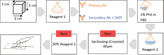
Methods: We modified the "clear unobstructed brain/body imaging cocktails" method, originally developed for whole-brain imaging in mice, and applied it to human colon tissue samples measuring approximately 10 mm3, a standard size typically processed in pathological laboratories.
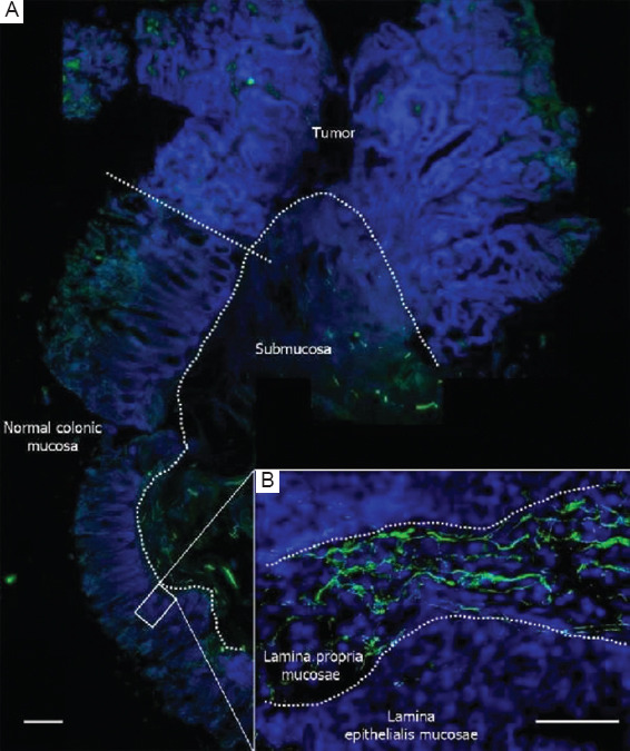
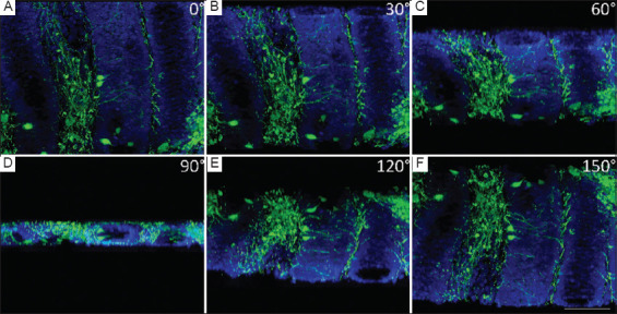
Results: Our protocol, which integrates a tissue-clearing technique, enabled reliable immunofluorescent visualization of colonic nerve fibers labeled with anti-β3-tubulin antibodies. The labeled nerve fibers could be observed using a standard epifluorescence microscope, and high-quality 3D reconstructions were generated through a simple image analysis approach using the open-source software ilastik, which eliminates the need for confocal microscopy.
Conclusion: The proposed steps provide a valuable method for researchers to visualize complex 3D structures, such as neural cells and processes, in both normal and tumor-transformed tissue settings.


 分享
分享

 求助内容:
求助内容: 应助结果提醒方式:
 扫码关注我们
