{"title":"The hazards of lack of co-registration of ictal brain SPECT with MRI: A case report of sinusitis mimicking a brainstem seizure focus.","authors":"Tracy Butler, Lawrence J Hirsch, Jan Claassen","doi":"10.1186/1471-2385-4-2","DOIUrl":null,"url":null,"abstract":"<p><p>BACKGROUND: Single photon emission computed tomography (SPECT) following injection of radiotracer during a seizure is known as ictal SPECT. Comparison of an ictal SPECT study to a baseline or interictal study can aid identification of a seizure focus. CASE PRESENTATION: A young woman with encephalitis and refractory seizures underwent brain SPECT during a period of frequent seizure-like episodes, and during a seizure-free period. A focal area of increased radiotracer uptake present only when she was experiencing frequent seizure-like episodes was originally localized to the brainstem, but with later computerized co-registration of SPECT to MRI, was found to lie outside the brain, in the region of the sphenoid sinus. CONCLUSION: Low-resolution SPECT images present difficulties in interpretation, which can be overcome through co-registration to higher-resolution structural images.</p>","PeriodicalId":80684,"journal":{"name":"BMC nuclear medicine","volume":"4 1","pages":"2"},"PeriodicalIF":0.0000,"publicationDate":"2004-11-29","publicationTypes":"Journal Article","fieldsOfStudy":null,"isOpenAccess":false,"openAccessPdf":"https://sci-hub-pdf.com/10.1186/1471-2385-4-2","citationCount":"3","resultStr":null,"platform":"Semanticscholar","paperid":null,"PeriodicalName":"BMC nuclear medicine","FirstCategoryId":"1085","ListUrlMain":"https://doi.org/10.1186/1471-2385-4-2","RegionNum":0,"RegionCategory":null,"ArticlePicture":[],"TitleCN":null,"AbstractTextCN":null,"PMCID":null,"EPubDate":"","PubModel":"","JCR":"","JCRName":"","Score":null,"Total":0}
引用次数: 3
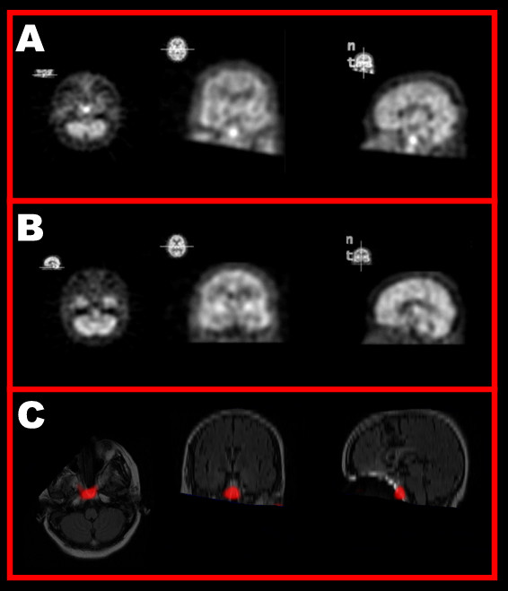
Abstract
BACKGROUND: Single photon emission computed tomography (SPECT) following injection of radiotracer during a seizure is known as ictal SPECT. Comparison of an ictal SPECT study to a baseline or interictal study can aid identification of a seizure focus. CASE PRESENTATION: A young woman with encephalitis and refractory seizures underwent brain SPECT during a period of frequent seizure-like episodes, and during a seizure-free period. A focal area of increased radiotracer uptake present only when she was experiencing frequent seizure-like episodes was originally localized to the brainstem, but with later computerized co-registration of SPECT to MRI, was found to lie outside the brain, in the region of the sphenoid sinus. CONCLUSION: Low-resolution SPECT images present difficulties in interpretation, which can be overcome through co-registration to higher-resolution structural images.


 分享
分享

 求助内容:
求助内容: 应助结果提醒方式:
 扫码关注我们
