Daniel O Pinto, Sarah Al Sharif, Gifty Mensah, Maria Cowen, Pooja Khatkar, James Erickson, Heather Branscome, Thomas Lattanze, Catherine DeMarino, Farhang Alem, Ruben Magni, Weidong Zhou, Sandrine Alais, Hélène Dutartre, Nazira El-Hage, Renaud Mahieux, Lance A Liotta, Fatah Kashanchi
{"title":"Extracellular vesicles from HTLV-1 infected cells modulate target cells and viral spread.","authors":"Daniel O Pinto, Sarah Al Sharif, Gifty Mensah, Maria Cowen, Pooja Khatkar, James Erickson, Heather Branscome, Thomas Lattanze, Catherine DeMarino, Farhang Alem, Ruben Magni, Weidong Zhou, Sandrine Alais, Hélène Dutartre, Nazira El-Hage, Renaud Mahieux, Lance A Liotta, Fatah Kashanchi","doi":"10.1186/s12977-021-00550-8","DOIUrl":null,"url":null,"abstract":"<p><strong>Background: </strong>The Human T-cell Lymphotropic Virus Type-1 (HTLV-1) is a blood-borne pathogen and etiological agent of Adult T-cell Leukemia/Lymphoma (ATLL) and HTLV-1 Associated Myelopathy/Tropical Spastic Paraparesis (HAM/TSP). HTLV-1 has currently infected up to 10 million globally with highly endemic areas in Japan, Africa, the Caribbean and South America. We have previously shown that Extracellular Vesicles (EVs) enhance HTLV-1 transmission by promoting cell-cell contact.</p><p><strong>Results: </strong>Here, we separated EVs into subpopulations using differential ultracentrifugation (DUC) at speeds of 2 k (2000×g), 10 k (10,000×g), and 100 k (100,000×g) from infected cell supernatants. Proteomic analysis revealed that EVs contain the highest viral/host protein abundance in the 2 k subpopulation (2 k > 10 k > 100 k). The 2 k and 10 k populations contained viral proteins (i.e., p19 and Tax), and autophagy proteins (i.e., LC3 and p62) suggesting presence of autophagosomes as well as core histones. Interestingly, the use of 2 k EVs in an angiogenesis assay (mesenchymal stem cells + endothelial cells) caused deterioration of vascular-like-tubules. Cells commonly associated with the neurovascular unit (i.e., astrocytes, neurons, and macrophages) in the blood-brain barrier (BBB) showed that HTLV-1 EVs may induce expression of cytokines involved in migration (i.e., IL-8; 100 k > 2 k > 10 k) from astrocytes and monocyte-derived macrophages (i.e., IL-8; 2 k > 10 k). Finally, we found that EVs were able to promote cell-cell contact and viral transmission in monocytic cell-derived dendritic cell. The EVs from both 2 k and 10 k increased HTLV-1 spread in a humanized mouse model, as evidenced by an increase in proviral DNA and RNA in the Blood, Lymph Node, and Spleen.</p><p><strong>Conclusions: </strong>Altogether, these data suggest that various EV subpopulations induce cytokine expression, tissue damage, and viral spread.</p>","PeriodicalId":2,"journal":{"name":"ACS Applied Bio Materials","volume":" ","pages":"6"},"PeriodicalIF":4.7000,"publicationDate":"2021-02-23","publicationTypes":"Journal Article","fieldsOfStudy":null,"isOpenAccess":false,"openAccessPdf":"https://www.ncbi.nlm.nih.gov/pmc/articles/PMC7901226/pdf/","citationCount":"0","resultStr":null,"platform":"Semanticscholar","paperid":null,"PeriodicalName":"ACS Applied Bio Materials","FirstCategoryId":"3","ListUrlMain":"https://doi.org/10.1186/s12977-021-00550-8","RegionNum":0,"RegionCategory":null,"ArticlePicture":[],"TitleCN":null,"AbstractTextCN":null,"PMCID":null,"EPubDate":"","PubModel":"","JCR":"Q2","JCRName":"MATERIALS SCIENCE, BIOMATERIALS","Score":null,"Total":0}
引用次数: 0
Abstract
Background: The Human T-cell Lymphotropic Virus Type-1 (HTLV-1) is a blood-borne pathogen and etiological agent of Adult T-cell Leukemia/Lymphoma (ATLL) and HTLV-1 Associated Myelopathy/Tropical Spastic Paraparesis (HAM/TSP). HTLV-1 has currently infected up to 10 million globally with highly endemic areas in Japan, Africa, the Caribbean and South America. We have previously shown that Extracellular Vesicles (EVs) enhance HTLV-1 transmission by promoting cell-cell contact.
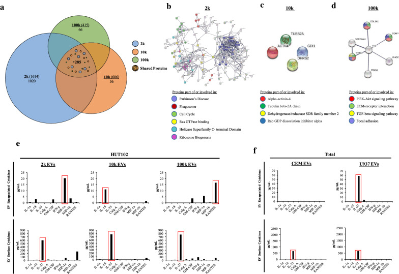
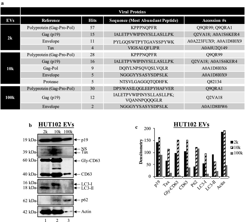
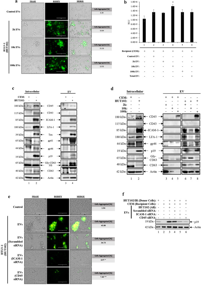
Results: Here, we separated EVs into subpopulations using differential ultracentrifugation (DUC) at speeds of 2 k (2000×g), 10 k (10,000×g), and 100 k (100,000×g) from infected cell supernatants. Proteomic analysis revealed that EVs contain the highest viral/host protein abundance in the 2 k subpopulation (2 k > 10 k > 100 k). The 2 k and 10 k populations contained viral proteins (i.e., p19 and Tax), and autophagy proteins (i.e., LC3 and p62) suggesting presence of autophagosomes as well as core histones. Interestingly, the use of 2 k EVs in an angiogenesis assay (mesenchymal stem cells + endothelial cells) caused deterioration of vascular-like-tubules. Cells commonly associated with the neurovascular unit (i.e., astrocytes, neurons, and macrophages) in the blood-brain barrier (BBB) showed that HTLV-1 EVs may induce expression of cytokines involved in migration (i.e., IL-8; 100 k > 2 k > 10 k) from astrocytes and monocyte-derived macrophages (i.e., IL-8; 2 k > 10 k). Finally, we found that EVs were able to promote cell-cell contact and viral transmission in monocytic cell-derived dendritic cell. The EVs from both 2 k and 10 k increased HTLV-1 spread in a humanized mouse model, as evidenced by an increase in proviral DNA and RNA in the Blood, Lymph Node, and Spleen.
Conclusions: Altogether, these data suggest that various EV subpopulations induce cytokine expression, tissue damage, and viral spread.
期刊介绍:
ACS Applied Bio Materials is an interdisciplinary journal publishing original research covering all aspects of biomaterials and biointerfaces including and beyond the traditional biosensing, biomedical and therapeutic applications.
The journal is devoted to reports of new and original experimental and theoretical research of an applied nature that integrates knowledge in the areas of materials, engineering, physics, bioscience, and chemistry into important bio applications. The journal is specifically interested in work that addresses the relationship between structure and function and assesses the stability and degradation of materials under relevant environmental and biological conditions.


 分享
分享
 求助内容:
求助内容: 应助结果提醒方式:
 扫码关注我们
