Anne E Tebo, Robert L Schmidt, Kamran Kadkhoda, Lisa K Peterson, Edward K L Chan, Marvin J Fritzler, Mark H Wener
{"title":"The antinuclear antibody HEp-2 indirect immunofluorescence assay: a survey of laboratory performance, pattern recognition and interpretation.","authors":"Anne E Tebo, Robert L Schmidt, Kamran Kadkhoda, Lisa K Peterson, Edward K L Chan, Marvin J Fritzler, Mark H Wener","doi":"10.1186/s13317-020-00146-w","DOIUrl":null,"url":null,"abstract":"<p><strong>Background: </strong>To evaluate the interpretation and reporting of antinuclear antibodies (ANA) by indirect immunofluorescence assay (IFA) using HEp-2 substrates based on common practice and guidance by the International Consensus on ANA patterns (ICAP).</p><p><strong>Method: </strong>Participants included two groups [16 clinical laboratories (CL) and 8 in vitro diagnostic manufacturers (IVD)] recruited via an email sent to the Association of Medical Laboratory Immunologists (AMLI) membership. Twelve (n = 12) pre-qualified specimens were distributed to participants for testing, interpretation and reporting HEp-2 IFA. Results obtained were analyzed for accuracy with the intended and consensus response for three main categorical patterns (nuclear, cytoplasmic and mitotic), common patterns and ICAP report nomenclatures. The distributions of antibody titers of specimens were also compared.</p><p><strong>Results: </strong>Laboratories differed in the categorical patterns reported; 8 reporting all patterns, 3 reporting only nuclear patterns and 5 reporting nuclear patterns with various combinations of other patterns. For all participants, accuracy with the intended response for the categorical nuclear pattern was excellent at 99% [95% confidence interval (CI): 97-100%] compared to 78% [95% CI 67-88%] for the cytoplasmic, and 93% [95% CI 86%-100%] for mitotic patterns. The accuracy was 13% greater for the common nomenclature [87%, 95% CI 82-90%] compared to the ICAP nomenclature [74%, 95% CI 68-79%] for all participants. Participants reporting all three main categories demonstrated better performances compared to those reporting 2 or less categorical patterns. The average accuracies varied between participant groups, however, with the lowest and most variable performances for cytoplasmic pattern specimens. The reported titers for all specimens varied, with the least variability for nuclear patterns and most titer variability associated with cytoplasmic patterns.</p><p><strong>Conclusions: </strong>Our study demonstrated significant accuracy for all participants in identifying the categorical nuclear staining as well as traditional pattern assignments for nuclear patterns. However, there was less consistency in reporting cytoplasmic and mitotic patterns, with implications for assigning competencies and training for clinical laboratory personnel.</p>","PeriodicalId":8655,"journal":{"name":"Auto-Immunity Highlights","volume":"12 1","pages":"4"},"PeriodicalIF":0.0000,"publicationDate":"2021-02-27","publicationTypes":"Journal Article","fieldsOfStudy":null,"isOpenAccess":false,"openAccessPdf":"https://sci-hub-pdf.com/10.1186/s13317-020-00146-w","citationCount":"5","resultStr":null,"platform":"Semanticscholar","paperid":null,"PeriodicalName":"Auto-Immunity Highlights","FirstCategoryId":"1085","ListUrlMain":"https://doi.org/10.1186/s13317-020-00146-w","RegionNum":0,"RegionCategory":null,"ArticlePicture":[],"TitleCN":null,"AbstractTextCN":null,"PMCID":null,"EPubDate":"","PubModel":"","JCR":"Q1","JCRName":"Medicine","Score":null,"Total":0}
引用次数: 5
Abstract
Background: To evaluate the interpretation and reporting of antinuclear antibodies (ANA) by indirect immunofluorescence assay (IFA) using HEp-2 substrates based on common practice and guidance by the International Consensus on ANA patterns (ICAP).
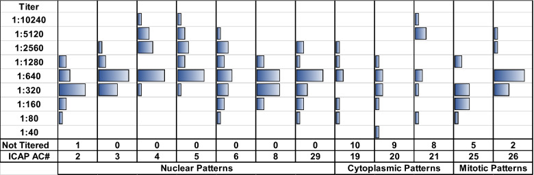
Method: Participants included two groups [16 clinical laboratories (CL) and 8 in vitro diagnostic manufacturers (IVD)] recruited via an email sent to the Association of Medical Laboratory Immunologists (AMLI) membership. Twelve (n = 12) pre-qualified specimens were distributed to participants for testing, interpretation and reporting HEp-2 IFA. Results obtained were analyzed for accuracy with the intended and consensus response for three main categorical patterns (nuclear, cytoplasmic and mitotic), common patterns and ICAP report nomenclatures. The distributions of antibody titers of specimens were also compared.
Results: Laboratories differed in the categorical patterns reported; 8 reporting all patterns, 3 reporting only nuclear patterns and 5 reporting nuclear patterns with various combinations of other patterns. For all participants, accuracy with the intended response for the categorical nuclear pattern was excellent at 99% [95% confidence interval (CI): 97-100%] compared to 78% [95% CI 67-88%] for the cytoplasmic, and 93% [95% CI 86%-100%] for mitotic patterns. The accuracy was 13% greater for the common nomenclature [87%, 95% CI 82-90%] compared to the ICAP nomenclature [74%, 95% CI 68-79%] for all participants. Participants reporting all three main categories demonstrated better performances compared to those reporting 2 or less categorical patterns. The average accuracies varied between participant groups, however, with the lowest and most variable performances for cytoplasmic pattern specimens. The reported titers for all specimens varied, with the least variability for nuclear patterns and most titer variability associated with cytoplasmic patterns.
Conclusions: Our study demonstrated significant accuracy for all participants in identifying the categorical nuclear staining as well as traditional pattern assignments for nuclear patterns. However, there was less consistency in reporting cytoplasmic and mitotic patterns, with implications for assigning competencies and training for clinical laboratory personnel.
背景:根据国际ANA模式共识(ICAP)的惯例和指导,评估使用HEp-2底物的间接免疫荧光测定(IFA)对抗核抗体(ANA)的解释和报告。方法:通过向医学实验室免疫学家协会(AMLI)会员发送电子邮件招募两组参与者[16家临床实验室(CL)和8家体外诊断制造商(IVD)]。12个(n = 12)预先合格的标本分发给参与者进行检测、解释和报告HEp-2 IFA。对获得的结果进行了准确性分析,并对三种主要分类模式(核、细胞质和有丝分裂)、常见模式和ICAP报告命名进行了预期和一致的反应。比较各标本的抗体滴度分布。结果:不同实验室报告的分类模式不同;8个报告所有模式,3个报告核模式,5个报告核模式与其他模式的各种组合。对于所有参与者,分类核模式预期反应的准确性为99%[95%置信区间(CI): 97-100%],而细胞质模式为78% [95% CI 67-88%],有丝分裂模式为93% [95% CI 86%-100%]。与ICAP命名法[74%,95% CI 68-79%]相比,所有参与者使用通用命名法的准确率要高13% [87%,95% CI 82-90%]。与报告两个或更少类别模式的参与者相比,报告所有三个主要类别的参与者表现出更好的表现。然而,参与者组之间的平均准确性各不相同,细胞质模式标本的表现最低,变化最大。所有标本报告的滴度各不相同,细胞核模式的变异最小,而大多数滴度变异与细胞质模式有关。结论:我们的研究证明了所有参与者在识别分类核染色以及核模式的传统模式分配方面的显著准确性。然而,报告细胞质和有丝分裂模式的一致性较差,这意味着临床实验室人员的能力分配和培训。


 分享
分享
 求助内容:
求助内容: 应助结果提醒方式:
 扫码关注我们
