Misty Ruppert, John Pyun, K V Chalam, David Sierpina
{"title":"Multimodal Imaging Characteristics of ADRP in a Family with p.Thr58Arg Substituted RHO Mutation.","authors":"Misty Ruppert, John Pyun, K V Chalam, David Sierpina","doi":"10.1155/2020/8860863","DOIUrl":null,"url":null,"abstract":"<p><strong>Background: </strong>Autosomal dominant retinitis pigmentosa (adRP) is a rare cause of progressive visual impairment in young patients and is frequently a result of RHO gene mutations. p.Thr58Arg rhodopsin mutation leads to misfolding of rhodopsin, subsequent accumulation in the endoplasmic reticulum, and leads to consecutive atrophy of photoreceptor cells through apoptosis.</p><p><strong>Materials and methods: </strong>We describe multimodal imaging findings in a 58-year-old female with adRP due to a c.173 C > G, p.Thr58Arg rhodopsin mutation (confirmed on genotyping), including ultra-wide-field fundus autofluorescence (UWF-FAF), color scanning laser ophthalmoscopy, structural optical coherence tomography (OCT), OCT-angiography (OCT-A), electroretinography (ERG), and visual field testing (HVF). Additionally, we compare the patient's phenotypic findings to those of her offspring, who was also affected by adRP.</p><p><strong>Results: </strong>The 58-year-old female and her son with symptoms of nyctalopia and decreased vision showed macular pigmentary changes in a bull's-eye pattern along with bone spicules in periphery with retinal atrophy. Genotyping confirmed p.Thr58Arg rhodopsin mutation. Wide area of dystrophic retina was noted on UWF-FAF, along with corresponding atrophy of photoreceptor layer on OCT. OCTA revealed complete nonperfusion of the superficial capillary plexus in areas of retinal dystrophy. ERG revealed increased latency and decreased amplitudes; HVF revealed constriction of visual fields consistent with retinal findings.</p><p><strong>Conclusions: </strong>Multimodal imaging is extremely helpful in delineating the extent of retinal dystrophy and comparable to ERG for monitoring of progress in retinitis pigmentosa. Photoreceptor layer thickness (measured with OCT) strongly correlated with ERG and can be used as a secondary surrogate for monitoring the disease progress.</p>","PeriodicalId":30325,"journal":{"name":"Case Reports in Genetics","volume":"2020 ","pages":"8860863"},"PeriodicalIF":0.0000,"publicationDate":"2020-12-02","publicationTypes":"Journal Article","fieldsOfStudy":null,"isOpenAccess":false,"openAccessPdf":"https://www.ncbi.nlm.nih.gov/pmc/articles/PMC7969344/pdf/","citationCount":"0","resultStr":null,"platform":"Semanticscholar","paperid":null,"PeriodicalName":"Case Reports in Genetics","FirstCategoryId":"1085","ListUrlMain":"https://doi.org/10.1155/2020/8860863","RegionNum":0,"RegionCategory":null,"ArticlePicture":[],"TitleCN":null,"AbstractTextCN":null,"PMCID":null,"EPubDate":"2020/1/1 0:00:00","PubModel":"eCollection","JCR":"","JCRName":"","Score":null,"Total":0}
引用次数: 0
Abstract
Background: Autosomal dominant retinitis pigmentosa (adRP) is a rare cause of progressive visual impairment in young patients and is frequently a result of RHO gene mutations. p.Thr58Arg rhodopsin mutation leads to misfolding of rhodopsin, subsequent accumulation in the endoplasmic reticulum, and leads to consecutive atrophy of photoreceptor cells through apoptosis.
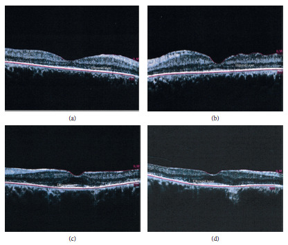
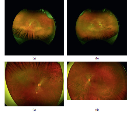
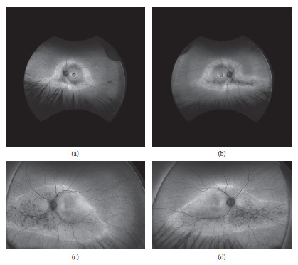
Materials and methods: We describe multimodal imaging findings in a 58-year-old female with adRP due to a c.173 C > G, p.Thr58Arg rhodopsin mutation (confirmed on genotyping), including ultra-wide-field fundus autofluorescence (UWF-FAF), color scanning laser ophthalmoscopy, structural optical coherence tomography (OCT), OCT-angiography (OCT-A), electroretinography (ERG), and visual field testing (HVF). Additionally, we compare the patient's phenotypic findings to those of her offspring, who was also affected by adRP.
Results: The 58-year-old female and her son with symptoms of nyctalopia and decreased vision showed macular pigmentary changes in a bull's-eye pattern along with bone spicules in periphery with retinal atrophy. Genotyping confirmed p.Thr58Arg rhodopsin mutation. Wide area of dystrophic retina was noted on UWF-FAF, along with corresponding atrophy of photoreceptor layer on OCT. OCTA revealed complete nonperfusion of the superficial capillary plexus in areas of retinal dystrophy. ERG revealed increased latency and decreased amplitudes; HVF revealed constriction of visual fields consistent with retinal findings.
Conclusions: Multimodal imaging is extremely helpful in delineating the extent of retinal dystrophy and comparable to ERG for monitoring of progress in retinitis pigmentosa. Photoreceptor layer thickness (measured with OCT) strongly correlated with ERG and can be used as a secondary surrogate for monitoring the disease progress.


 分享
分享

 求助内容:
求助内容: 应助结果提醒方式:
 扫码关注我们
