{"title":"Muscle atrophy induced by overexpression of ALAS2 is related to muscle mitochondrial dysfunction.","authors":"Yahui Peng, Jihong Li, Dixian Luo, Shuai Zhang, Sijia Li, Dayong Wang, Xidi Wang, Zhujun Zhang, Xue Wang, Changhui Sun, Xu Gao, Yang Hui, Rongzhang He","doi":"10.1186/s13395-021-00263-8","DOIUrl":null,"url":null,"abstract":"<p><strong>Background: </strong>ALAS2 (delta-aminolevulinate synthase 2) is one of the two isoenzymes catalyzing the synthesis of delta-aminolevulinic acid (ALA), which is the first precursor of heme synthesis. ALAS2-overexpressing transgenic mice (Tg mice) showed syndrome of porphyria, a series of diseases related to the heme anabolism deficiency. Tg mice showed an obvious decrease in muscle size. Muscle atrophy results from a decrease in protein synthesis and an increase in protein degradation, which ultimately leads to a decrease in myofiber size due to loss of contractile proteins, organelles, nuclei, and cytoplasm.</p><p><strong>Methods: </strong>The forelimb muscle grip strength of age-matched ALAS-2 transgenic mice (Tg mice) and wild-type mice (WT mice) were measured with an automated grip strength meter. The activities of serum LDH and CK-MB were measured by Modular DPP. The histology of skeletal muscle (quadriceps femoris and gastrocnemius) was observed by hematoxylin and eosin (HE) staining, immunohistochemistry, and transmission electron microscope. Real-time PCR was used to detect mtDNA content and UCP3 mRNA expression. Evans blue dye staining was used to detect the membrane damage of the muscle fiber. Single skeletal muscle fiber diameter was measured by single-fiber analyses. Muscle adenosine triphosphate (ATP) levels were detected by a luminometric assay with an ATP assay kit.</p><p><strong>Results: </strong>Compared with WT mice, the strength of forelimb muscle and mass of gastrocnemius were decreased in Tg mice. The activities of serum CK-MB and LDH, the number of central nuclei fibers, and Evans blue positive fibers were more than those in WT mice, while the diameter of single fibers was smaller, which were associated with suppressed expression levels of MHC, myoD1, dystrophin, atrogin1, and MuRF1. Re-expression of eMyHC was only showed in the quadriceps of Tg mice, but not in WT mice. Muscle mitochondria in Tg mice showed dysfunction with descented ATP production and mtDNA content, downregulated UCP3 mRNA expression, and swelling of mitochondria.</p><p><strong>Conclusion: </strong>ALAS2 overexpressing-transgenic mice (Tg mice) showed muscle dystrophy, which was associated with decreased atrogin-1 and MuRF-1, and closely related to mitochondrial dysfunction.</p>","PeriodicalId":21747,"journal":{"name":"Skeletal Muscle","volume":" ","pages":"9"},"PeriodicalIF":4.4000,"publicationDate":"2021-03-30","publicationTypes":"Journal Article","fieldsOfStudy":null,"isOpenAccess":false,"openAccessPdf":"https://sci-hub-pdf.com/10.1186/s13395-021-00263-8","citationCount":"6","resultStr":null,"platform":"Semanticscholar","paperid":null,"PeriodicalName":"Skeletal Muscle","FirstCategoryId":"3","ListUrlMain":"https://doi.org/10.1186/s13395-021-00263-8","RegionNum":2,"RegionCategory":"医学","ArticlePicture":[],"TitleCN":null,"AbstractTextCN":null,"PMCID":null,"EPubDate":"","PubModel":"","JCR":"Q2","JCRName":"CELL BIOLOGY","Score":null,"Total":0}
引用次数: 6
Abstract
Background: ALAS2 (delta-aminolevulinate synthase 2) is one of the two isoenzymes catalyzing the synthesis of delta-aminolevulinic acid (ALA), which is the first precursor of heme synthesis. ALAS2-overexpressing transgenic mice (Tg mice) showed syndrome of porphyria, a series of diseases related to the heme anabolism deficiency. Tg mice showed an obvious decrease in muscle size. Muscle atrophy results from a decrease in protein synthesis and an increase in protein degradation, which ultimately leads to a decrease in myofiber size due to loss of contractile proteins, organelles, nuclei, and cytoplasm.
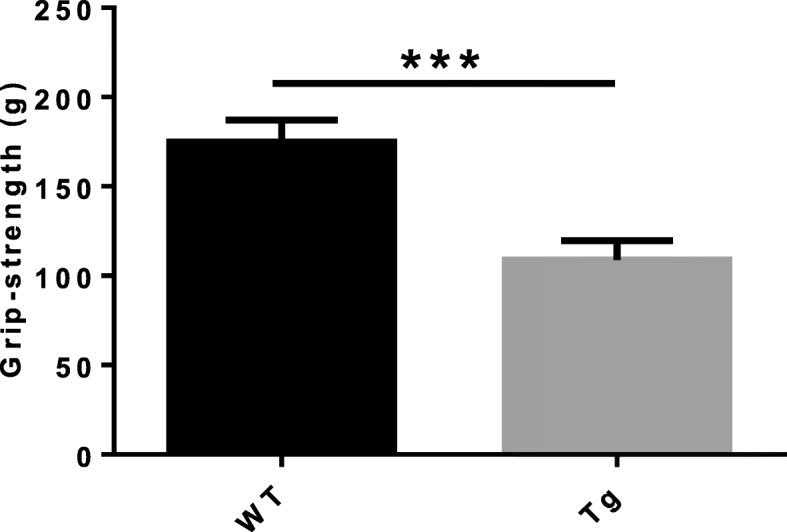
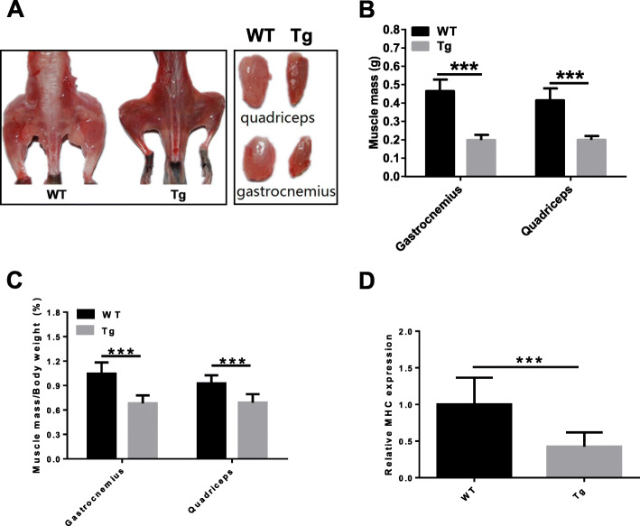
Methods: The forelimb muscle grip strength of age-matched ALAS-2 transgenic mice (Tg mice) and wild-type mice (WT mice) were measured with an automated grip strength meter. The activities of serum LDH and CK-MB were measured by Modular DPP. The histology of skeletal muscle (quadriceps femoris and gastrocnemius) was observed by hematoxylin and eosin (HE) staining, immunohistochemistry, and transmission electron microscope. Real-time PCR was used to detect mtDNA content and UCP3 mRNA expression. Evans blue dye staining was used to detect the membrane damage of the muscle fiber. Single skeletal muscle fiber diameter was measured by single-fiber analyses. Muscle adenosine triphosphate (ATP) levels were detected by a luminometric assay with an ATP assay kit.
Results: Compared with WT mice, the strength of forelimb muscle and mass of gastrocnemius were decreased in Tg mice. The activities of serum CK-MB and LDH, the number of central nuclei fibers, and Evans blue positive fibers were more than those in WT mice, while the diameter of single fibers was smaller, which were associated with suppressed expression levels of MHC, myoD1, dystrophin, atrogin1, and MuRF1. Re-expression of eMyHC was only showed in the quadriceps of Tg mice, but not in WT mice. Muscle mitochondria in Tg mice showed dysfunction with descented ATP production and mtDNA content, downregulated UCP3 mRNA expression, and swelling of mitochondria.
Conclusion: ALAS2 overexpressing-transgenic mice (Tg mice) showed muscle dystrophy, which was associated with decreased atrogin-1 and MuRF-1, and closely related to mitochondrial dysfunction.
期刊介绍:
The only open access journal in its field, Skeletal Muscle publishes novel, cutting-edge research and technological advancements that investigate the molecular mechanisms underlying the biology of skeletal muscle. Reflecting the breadth of research in this area, the journal welcomes manuscripts about the development, metabolism, the regulation of mass and function, aging, degeneration, dystrophy and regeneration of skeletal muscle, with an emphasis on understanding adult skeletal muscle, its maintenance, and its interactions with non-muscle cell types and regulatory modulators.
Main areas of interest include:
-differentiation of skeletal muscle-
atrophy and hypertrophy of skeletal muscle-
aging of skeletal muscle-
regeneration and degeneration of skeletal muscle-
biology of satellite and satellite-like cells-
dystrophic degeneration of skeletal muscle-
energy and glucose homeostasis in skeletal muscle-
non-dystrophic genetic diseases of skeletal muscle, such as Spinal Muscular Atrophy and myopathies-
maintenance of neuromuscular junctions-
roles of ryanodine receptors and calcium signaling in skeletal muscle-
roles of nuclear receptors in skeletal muscle-
roles of GPCRs and GPCR signaling in skeletal muscle-
other relevant aspects of skeletal muscle biology.
In addition, articles on translational clinical studies that address molecular and cellular mechanisms of skeletal muscle will be published. Case reports are also encouraged for submission.
Skeletal Muscle reflects the breadth of research on skeletal muscle and bridges gaps between diverse areas of science for example cardiac cell biology and neurobiology, which share common features with respect to cell differentiation, excitatory membranes, cell-cell communication, and maintenance. Suitable articles are model and mechanism-driven, and apply statistical principles where appropriate; purely descriptive studies are of lesser interest.


 分享
分享
 求助内容:
求助内容: 应助结果提醒方式:
 扫码关注我们
