{"title":"Atlas of sentinel lymph nodes in early breast cancer using single-photon emission computed tomography: implication for lymphatic contouring.","authors":"Sergey Nikolaevich Novikov, Pavel Ivanovich Krzhivitskii, Yulia Sergeevna Melnik, Alina Albertovna Valitova, Zhanna Viktorovna Bryantseva, Irina Alexandrovna Akulova, Sergey Vasilevich Kanaev","doi":"10.3857/roj.2020.00871","DOIUrl":null,"url":null,"abstract":"<p><strong>Purpose: </strong>to determine the localization of sentinel lymph nodes (SLNs) in a large cohort of patients with breast cancer and validate the European Society for Therapeutic Radiology and Oncology (ESTRO), Radiation Therapy Oncology Group (RTOG), and Radiotherapy Comparative Effectiveness (RADCOMP) guidelines on regional lymph node clinical target volume (CTV-LN) delineation.</p><p><strong>Materials and methods: </strong>A total of 254 women with cT1-3N0-1M0 breast cancer underwent single-photon emission computed tomography (SPECT-CT) visualization of SLNs after intra- and peritumoral injection of 99mTc-radiocolloids. All SPECT-CT images were fused with reference simulation computed tomography. A 3D atlas of SLNs was created and used for evaluation of CTV-LN defined by contouring guidelines.</p><p><strong>Results: </strong>SPECT-CT visualized 532 SLNs that were localized in axillary level I in 67.5%, level II in 15.4%, level III in 7.3%, internal mammary in 8.5%, and supraclavicular in 1.3% cases. The majority of level II-IV and internal mammary SLNs were inside the recommended CTV-LN. Axillary level I SLNs were covered by ESTRO and RTOG contours in 85% and 85% cases, respectively. \"Out of contours\" SLNs were mostly detected in lateral subgroup of level I LN (18.5%), while 98%-99% of anterior pectoral and central axillary SLNs were covered by CTV-LN. Internal mammary SLNs were visualized in 33 cases and were outside ESTRO and RTOG contours in 3 and 6 observations, respectively.</p><p><strong>Conclusion: </strong>SPECT-CT atlas of SLNs demonstrated that in most cases ESTRO and RTOG guidelines correctly represented CTV-LNs with the exception of lateral subgroup of SLNs.</p>","PeriodicalId":46572,"journal":{"name":"Radiation Oncology Journal","volume":"39 1","pages":"8-14"},"PeriodicalIF":2.2000,"publicationDate":"2021-03-01","publicationTypes":"Journal Article","fieldsOfStudy":null,"isOpenAccess":false,"openAccessPdf":"https://ftp.ncbi.nlm.nih.gov/pub/pmc/oa_pdf/26/ce/roj-2020-00871.PMC8024181.pdf","citationCount":"3","resultStr":null,"platform":"Semanticscholar","paperid":null,"PeriodicalName":"Radiation Oncology Journal","FirstCategoryId":"1085","ListUrlMain":"https://doi.org/10.3857/roj.2020.00871","RegionNum":0,"RegionCategory":null,"ArticlePicture":[],"TitleCN":null,"AbstractTextCN":null,"PMCID":null,"EPubDate":"2021/3/25 0:00:00","PubModel":"Epub","JCR":"Q3","JCRName":"ONCOLOGY","Score":null,"Total":0}
引用次数: 3
Abstract
Purpose: to determine the localization of sentinel lymph nodes (SLNs) in a large cohort of patients with breast cancer and validate the European Society for Therapeutic Radiology and Oncology (ESTRO), Radiation Therapy Oncology Group (RTOG), and Radiotherapy Comparative Effectiveness (RADCOMP) guidelines on regional lymph node clinical target volume (CTV-LN) delineation.
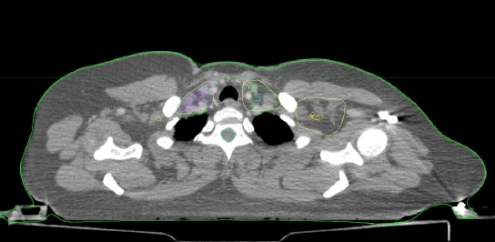
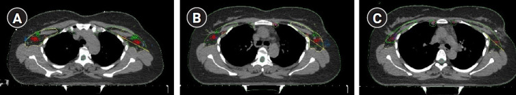
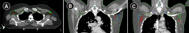
Materials and methods: A total of 254 women with cT1-3N0-1M0 breast cancer underwent single-photon emission computed tomography (SPECT-CT) visualization of SLNs after intra- and peritumoral injection of 99mTc-radiocolloids. All SPECT-CT images were fused with reference simulation computed tomography. A 3D atlas of SLNs was created and used for evaluation of CTV-LN defined by contouring guidelines.
Results: SPECT-CT visualized 532 SLNs that were localized in axillary level I in 67.5%, level II in 15.4%, level III in 7.3%, internal mammary in 8.5%, and supraclavicular in 1.3% cases. The majority of level II-IV and internal mammary SLNs were inside the recommended CTV-LN. Axillary level I SLNs were covered by ESTRO and RTOG contours in 85% and 85% cases, respectively. "Out of contours" SLNs were mostly detected in lateral subgroup of level I LN (18.5%), while 98%-99% of anterior pectoral and central axillary SLNs were covered by CTV-LN. Internal mammary SLNs were visualized in 33 cases and were outside ESTRO and RTOG contours in 3 and 6 observations, respectively.
Conclusion: SPECT-CT atlas of SLNs demonstrated that in most cases ESTRO and RTOG guidelines correctly represented CTV-LNs with the exception of lateral subgroup of SLNs.


 分享
分享
 求助内容:
求助内容: 应助结果提醒方式:
 扫码关注我们
