Fengfeng L Xu, Youssef Rbaibi, Kirill Kiselyov, John S Lazo, Peter Wipf, William S Saunders
{"title":"Mitotic slippage in non-cancer cells induced by a microtubule disruptor, disorazole C1.","authors":"Fengfeng L Xu, Youssef Rbaibi, Kirill Kiselyov, John S Lazo, Peter Wipf, William S Saunders","doi":"10.1186/1472-6769-10-1","DOIUrl":null,"url":null,"abstract":"<p><strong>Background: </strong>Disorazoles are polyene macrodiolides isolated from a myxobacterium fermentation broth. Disorazole C1 was newly synthesized and found to depolymerize microtubules and cause mitotic arrest. Here we examined the cellular responses to disorazole C1 in both non-cancer and cancer cells and compared our results to vinblastine and taxol.</p><p><strong>Results: </strong>In non-cancer cells, disorazole C1 induced a prolonged mitotic arrest, followed by mitotic slippage, as confirmed by live cell imaging and cell cycle analysis. This mitotic slippage was associated with cyclin B degradation, but did not require p53. Four assays for apoptosis, including western blotting for poly(ADP-ribose) polymerase cleavage, microscopic analyses for cytochrome C release and annexin V staining, and gel electrophoresis examination for DNA laddering, were conducted and demonstrated little induction of apoptosis in non-cancer cells treated with disorazole C1. On the contrary, we observed an activated apoptotic pathway in cancer cells, suggesting that normal and malignant cells respond differently to disorazole C1.</p><p><strong>Conclusion: </strong>Our studies demonstrate that non-cancer cells undergo mitotic slippage in a cyclin B-dependent and p53-independent manner after prolonged mitotic arrest caused by disorazole C1. In contrast, cancer cells induce the apoptotic pathway after disorazole C1 treatment, indicating a possibly significant therapeutic window for this compound.</p>","PeriodicalId":80682,"journal":{"name":"BMC chemical biology","volume":"10 ","pages":"1"},"PeriodicalIF":0.0000,"publicationDate":"2010-02-11","publicationTypes":"Journal Article","fieldsOfStudy":null,"isOpenAccess":false,"openAccessPdf":"https://sci-hub-pdf.com/10.1186/1472-6769-10-1","citationCount":"16","resultStr":null,"platform":"Semanticscholar","paperid":null,"PeriodicalName":"BMC chemical biology","FirstCategoryId":"1085","ListUrlMain":"https://doi.org/10.1186/1472-6769-10-1","RegionNum":0,"RegionCategory":null,"ArticlePicture":[],"TitleCN":null,"AbstractTextCN":null,"PMCID":null,"EPubDate":"","PubModel":"","JCR":"","JCRName":"","Score":null,"Total":0}
引用次数: 16
Abstract
Background: Disorazoles are polyene macrodiolides isolated from a myxobacterium fermentation broth. Disorazole C1 was newly synthesized and found to depolymerize microtubules and cause mitotic arrest. Here we examined the cellular responses to disorazole C1 in both non-cancer and cancer cells and compared our results to vinblastine and taxol.
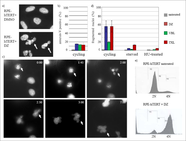
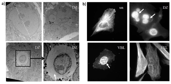
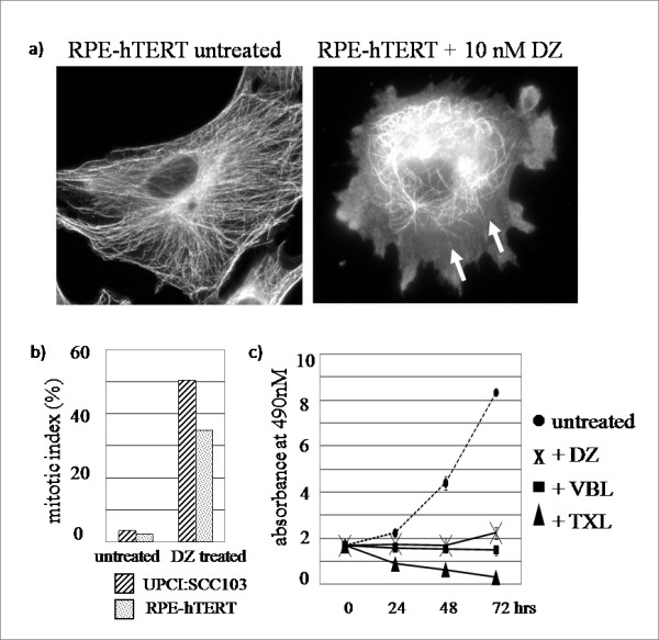
Results: In non-cancer cells, disorazole C1 induced a prolonged mitotic arrest, followed by mitotic slippage, as confirmed by live cell imaging and cell cycle analysis. This mitotic slippage was associated with cyclin B degradation, but did not require p53. Four assays for apoptosis, including western blotting for poly(ADP-ribose) polymerase cleavage, microscopic analyses for cytochrome C release and annexin V staining, and gel electrophoresis examination for DNA laddering, were conducted and demonstrated little induction of apoptosis in non-cancer cells treated with disorazole C1. On the contrary, we observed an activated apoptotic pathway in cancer cells, suggesting that normal and malignant cells respond differently to disorazole C1.
Conclusion: Our studies demonstrate that non-cancer cells undergo mitotic slippage in a cyclin B-dependent and p53-independent manner after prolonged mitotic arrest caused by disorazole C1. In contrast, cancer cells induce the apoptotic pathway after disorazole C1 treatment, indicating a possibly significant therapeutic window for this compound.


 分享
分享

 求助内容:
求助内容: 应助结果提醒方式:
 扫码关注我们
