Debra A O'Leary, Leonardo Vargas, Orzala Sharif, Michael E Garcia, Yury J Sigal, Siu-Kei Chow, Christian Schmedt, Jeremy S Caldwell, Achim Brinker, Ingo H Engels
{"title":"HTS-Compatible Patient-Derived Cell-Based Assay to Identify Small Molecule Modulators of Aberrant Splicing in Myotonic Dystrophy Type 1.","authors":"Debra A O'Leary, Leonardo Vargas, Orzala Sharif, Michael E Garcia, Yury J Sigal, Siu-Kei Chow, Christian Schmedt, Jeremy S Caldwell, Achim Brinker, Ingo H Engels","doi":"10.2174/1875397301004010009","DOIUrl":null,"url":null,"abstract":"<p><p>Myotonic dystrophy type 1 (DM1) is a genetic disorder characterized by muscle wasting, myotonia, cataracts, cardiac arrhythmia, hyperinsulinism and intellectual deficits, and is caused by expansion of a CTG repeat in the 3'UTR of the Dystrophia Myotonica-Protein Kinase (DMPK) gene. The DMPK transcripts containing expanded CUG repeats accumulate in nuclear foci and ultimately cause mis-splicing of secondary genes through the dysregulation of RNA-binding proteins including Muscleblind 1 (MBNL1) and CUG binding protein 1 (CUGBP1). Correction of mis-splicing of genes such as the Skeletal muscle-specific chloride channel 1 (CLCN1), Cardiac troponin T (TNNT2), Insulin receptor (INSR) and Sarcoplasmic/endoplasmic reticulum Ca(2+)ATPase 1 (SERCA1) may alleviate some of the symptoms of DM1; hence identification of small molecule modulators is an important step towards a therapy for DM1 patients. Here we describe the generation of immortalized myoblast cell lines derived from healthy (DMPK CTG(5)) and DM1 patient (DMPK CTG(1000)) fibroblasts by constitutive overexpression of human telomerase reverse transcriptase (hTERT) and inducible overexpression of the Myoblast determination factor (MYOD). MBNL1-containing nuclear foci, mis-splicing events and defective myotube differentiation defects characteristic of DM1 were observed in these cells. A CLCN1 luciferase minigene construct (CLCN1-luc) was stably introduced to monitor intron 2 retention in the DM1 cellular context (a reported splicing defect in DM1). The assay was validated by performing a high-throughput screen (HTS) of ~13,000 low molecular weight compounds against the CLCN1-luc DM1 myoblast cell line, providing an ideal system for conducting HTS to better understand and treat DM1.</p>","PeriodicalId":88232,"journal":{"name":"Current chemical genomics","volume":"4 ","pages":"9-18"},"PeriodicalIF":0.0000,"publicationDate":"2010-03-19","publicationTypes":"Journal Article","fieldsOfStudy":null,"isOpenAccess":false,"openAccessPdf":"https://ftp.ncbi.nlm.nih.gov/pub/pmc/oa_pdf/6e/db/TOCHGENJ-4-9.PMC2874217.pdf","citationCount":"19","resultStr":null,"platform":"Semanticscholar","paperid":null,"PeriodicalName":"Current chemical genomics","FirstCategoryId":"1085","ListUrlMain":"https://doi.org/10.2174/1875397301004010009","RegionNum":0,"RegionCategory":null,"ArticlePicture":[],"TitleCN":null,"AbstractTextCN":null,"PMCID":null,"EPubDate":"","PubModel":"","JCR":"","JCRName":"","Score":null,"Total":0}
引用次数: 19
Abstract
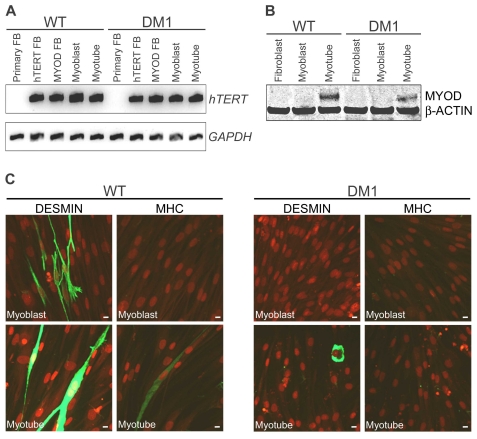
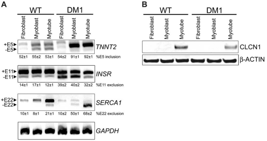
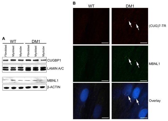
Myotonic dystrophy type 1 (DM1) is a genetic disorder characterized by muscle wasting, myotonia, cataracts, cardiac arrhythmia, hyperinsulinism and intellectual deficits, and is caused by expansion of a CTG repeat in the 3'UTR of the Dystrophia Myotonica-Protein Kinase (DMPK) gene. The DMPK transcripts containing expanded CUG repeats accumulate in nuclear foci and ultimately cause mis-splicing of secondary genes through the dysregulation of RNA-binding proteins including Muscleblind 1 (MBNL1) and CUG binding protein 1 (CUGBP1). Correction of mis-splicing of genes such as the Skeletal muscle-specific chloride channel 1 (CLCN1), Cardiac troponin T (TNNT2), Insulin receptor (INSR) and Sarcoplasmic/endoplasmic reticulum Ca(2+)ATPase 1 (SERCA1) may alleviate some of the symptoms of DM1; hence identification of small molecule modulators is an important step towards a therapy for DM1 patients. Here we describe the generation of immortalized myoblast cell lines derived from healthy (DMPK CTG(5)) and DM1 patient (DMPK CTG(1000)) fibroblasts by constitutive overexpression of human telomerase reverse transcriptase (hTERT) and inducible overexpression of the Myoblast determination factor (MYOD). MBNL1-containing nuclear foci, mis-splicing events and defective myotube differentiation defects characteristic of DM1 were observed in these cells. A CLCN1 luciferase minigene construct (CLCN1-luc) was stably introduced to monitor intron 2 retention in the DM1 cellular context (a reported splicing defect in DM1). The assay was validated by performing a high-throughput screen (HTS) of ~13,000 low molecular weight compounds against the CLCN1-luc DM1 myoblast cell line, providing an ideal system for conducting HTS to better understand and treat DM1.


 分享
分享

 求助内容:
求助内容: 应助结果提醒方式:
 扫码关注我们
