Percutaneous ilioinguinal-iliohypogastric nerve block or step-by-step local infiltration anesthesia for inguinal hernia repair: what cadaveric dissection says?
Hakan Kulacoglu, Zafer Ergul, Ali Firat Esmer, Tulin Sen, Taylan Akkaya, Alaittin Elhan
{"title":"Percutaneous ilioinguinal-iliohypogastric nerve block or step-by-step local infiltration anesthesia for inguinal hernia repair: what cadaveric dissection says?","authors":"Hakan Kulacoglu, Zafer Ergul, Ali Firat Esmer, Tulin Sen, Taylan Akkaya, Alaittin Elhan","doi":"10.4174/jkss.2011.81.6.408","DOIUrl":null,"url":null,"abstract":"<p><strong>Purpose: </strong>The repair of groin hernias with local anesthesia has gained popularity. Two main methods have been described for local anesthesia. This study was aimed at comparing percutaneous truncular ilioinguinal-iliohypogastric block and step-by-step infiltration technique by using cadaver dissections.</p><p><strong>Methods: </strong>The study was performed on an adult male cadaver by using blue dye injection. A percutaneous nerve block simulation was done on right side and the dye was given in between the internal oblique and transversus muscles. On the left side, a skin incision was deepened and the dye was injected under the external oblique aponeurosis. Following the injections, stained areas were investigated superficially and within the deeper tissues with dissection.</p><p><strong>Results: </strong>There was a complete superficial staining covering the iliohypogastric and ilioinguinal nerves in the inguinal floor at both sides. On the right side, intraabdominal observation showed a wide and intense peritoneal staining, while almost no staining was seen on the left side. Preperitoneal dissection displayed a massive staining including testicular vascular pedicule and vas deferens on the right side. The dye solution also infiltrated the area of the femoral nerve prominently. On the contrary, a very limited staining was seen on the left.</p><p><strong>Conclusion: </strong>It may not always be easy to keep the percutaneous block within optimum anatomical limits without causing adverse events. A step-by-step infiltration technique under direct surgical vision seems to be safer than percutaneous inguinal block for patients undergoing inguinal hernia repair.</p>","PeriodicalId":49157,"journal":{"name":"Journal of the Korean Surgical Society","volume":"81 6","pages":"408-13"},"PeriodicalIF":0.0000,"publicationDate":"2011-12-01","publicationTypes":"Journal Article","fieldsOfStudy":null,"isOpenAccess":false,"openAccessPdf":"https://sci-hub-pdf.com/10.4174/jkss.2011.81.6.408","citationCount":"8","resultStr":null,"platform":"Semanticscholar","paperid":null,"PeriodicalName":"Journal of the Korean Surgical Society","FirstCategoryId":"1085","ListUrlMain":"https://doi.org/10.4174/jkss.2011.81.6.408","RegionNum":0,"RegionCategory":null,"ArticlePicture":[],"TitleCN":null,"AbstractTextCN":null,"PMCID":null,"EPubDate":"2011/11/25 0:00:00","PubModel":"Epub","JCR":"","JCRName":"","Score":null,"Total":0}
引用次数: 8
Abstract
Purpose: The repair of groin hernias with local anesthesia has gained popularity. Two main methods have been described for local anesthesia. This study was aimed at comparing percutaneous truncular ilioinguinal-iliohypogastric block and step-by-step infiltration technique by using cadaver dissections.
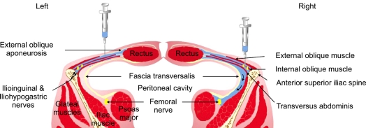
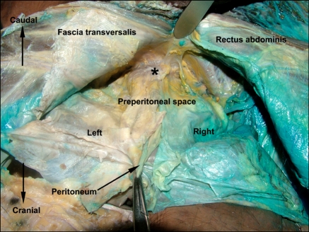
Methods: The study was performed on an adult male cadaver by using blue dye injection. A percutaneous nerve block simulation was done on right side and the dye was given in between the internal oblique and transversus muscles. On the left side, a skin incision was deepened and the dye was injected under the external oblique aponeurosis. Following the injections, stained areas were investigated superficially and within the deeper tissues with dissection.
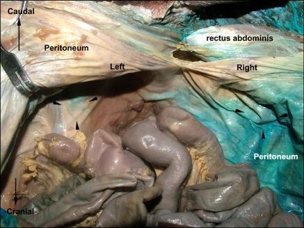
Results: There was a complete superficial staining covering the iliohypogastric and ilioinguinal nerves in the inguinal floor at both sides. On the right side, intraabdominal observation showed a wide and intense peritoneal staining, while almost no staining was seen on the left side. Preperitoneal dissection displayed a massive staining including testicular vascular pedicule and vas deferens on the right side. The dye solution also infiltrated the area of the femoral nerve prominently. On the contrary, a very limited staining was seen on the left.
Conclusion: It may not always be easy to keep the percutaneous block within optimum anatomical limits without causing adverse events. A step-by-step infiltration technique under direct surgical vision seems to be safer than percutaneous inguinal block for patients undergoing inguinal hernia repair.


 分享
分享
 求助内容:
求助内容: 应助结果提醒方式:
 扫码关注我们
