Ivana Joksic, Dragana Vujic, Marija Guc-Scekic, Andreja Leskovac, Sandra Petrovic, Maryam Ojani, Juan P Trujillo, Jordi Surralles, Maja Zivkovic, Aleksandra Stankovic, Predrag Slijepcevic, Gordana Joksic
{"title":"Dysfunctional telomeres in primary cells from Fanconi anemia FANCD2 patients.","authors":"Ivana Joksic, Dragana Vujic, Marija Guc-Scekic, Andreja Leskovac, Sandra Petrovic, Maryam Ojani, Juan P Trujillo, Jordi Surralles, Maja Zivkovic, Aleksandra Stankovic, Predrag Slijepcevic, Gordana Joksic","doi":"10.1186/2041-9414-3-6","DOIUrl":null,"url":null,"abstract":"<p><strong>Unlabelled: </strong></p><p><strong>Background: </strong>Fanconi anemia (FA) is characterized by sensitivity to DNA cross-linking agents, mild cellular, and marked clinical radio sensitivity. In this study we investigated telomeric abnormalities of non-immortalized primary cells (lymphocytes and fibroblasts) derived from FA patients of the FA-D2 complementation group, which provides a more accurate physiological assessment than is possible with transformed cells or animal models.</p><p><strong>Results: </strong>We analyzed telomere length, telomere dysfunction-induced foci (TIFs), sister chromatid exchanges (SCE), telomere sister chromatid exchanges (T-SCE), apoptosis and expression of shelterin components TRF1 and TRF2. FANCD2 lymphocytes exhibited multiple types of telomeric abnormalities, including premature telomere shortening, increase in telomeric recombination and aberrant telomeric structures ranging from fragile to long-string extended telomeres. The baseline incidence of SCE in FANCD2 lymphocytes was reduced when compared to control, but in response to diepoxybutane (DEB) the 2-fold higher rate of SCE was observed. In contrast, control lymphocytes showed decreased SCE incidence in response to DEB treatment. FANCD2 fibroblasts revealed a high percentage of TIFs, decreased expression of TRF1 and invariable expression of TRF2. The percentage of TIFs inversely correlated with telomere length, emphasizing that telomere shortening is the major reason for the loss of telomere capping function. Upon irradiation, a significant decrease of TIFs was observed at all recovery times. Surprisingly, a considerable percentage of TIF positive cells disappeared at the same time when incidence of γ-H2AX foci was maximal. Both FANCD2 leucocytes and fibroblasts appeared to die spontaneously at higher rate than control. This trend was more evident upon irradiation; the percentage of leucocytes underwent apoptosis was 2.59- fold higher than that in control, while fibroblasts exhibited a 2- h delay before entering apoptosis.</p><p><strong>Conclusion: </strong>The results of our study showed that primary cells originating from FA-D2 patients display shorten telomeres, elevated incidence of T-SCEs and high frequency of TIFs. Disappearance of TIFs in early response to irradiation represent distinctive feature of FANCD2 cells that should be examined further.</p>","PeriodicalId":53596,"journal":{"name":"Genome Integrity","volume":"3 1","pages":"6"},"PeriodicalIF":0.0000,"publicationDate":"2012-09-14","publicationTypes":"Journal Article","fieldsOfStudy":null,"isOpenAccess":false,"openAccessPdf":"https://sci-hub-pdf.com/10.1186/2041-9414-3-6","citationCount":"37","resultStr":null,"platform":"Semanticscholar","paperid":null,"PeriodicalName":"Genome Integrity","FirstCategoryId":"1085","ListUrlMain":"https://doi.org/10.1186/2041-9414-3-6","RegionNum":0,"RegionCategory":null,"ArticlePicture":[],"TitleCN":null,"AbstractTextCN":null,"PMCID":null,"EPubDate":"","PubModel":"","JCR":"Q4","JCRName":"Biochemistry, Genetics and Molecular Biology","Score":null,"Total":0}
引用次数: 37
Abstract
Unlabelled:
Background: Fanconi anemia (FA) is characterized by sensitivity to DNA cross-linking agents, mild cellular, and marked clinical radio sensitivity. In this study we investigated telomeric abnormalities of non-immortalized primary cells (lymphocytes and fibroblasts) derived from FA patients of the FA-D2 complementation group, which provides a more accurate physiological assessment than is possible with transformed cells or animal models.
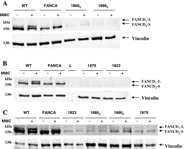
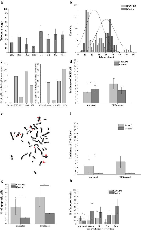
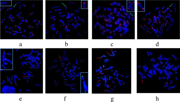
Results: We analyzed telomere length, telomere dysfunction-induced foci (TIFs), sister chromatid exchanges (SCE), telomere sister chromatid exchanges (T-SCE), apoptosis and expression of shelterin components TRF1 and TRF2. FANCD2 lymphocytes exhibited multiple types of telomeric abnormalities, including premature telomere shortening, increase in telomeric recombination and aberrant telomeric structures ranging from fragile to long-string extended telomeres. The baseline incidence of SCE in FANCD2 lymphocytes was reduced when compared to control, but in response to diepoxybutane (DEB) the 2-fold higher rate of SCE was observed. In contrast, control lymphocytes showed decreased SCE incidence in response to DEB treatment. FANCD2 fibroblasts revealed a high percentage of TIFs, decreased expression of TRF1 and invariable expression of TRF2. The percentage of TIFs inversely correlated with telomere length, emphasizing that telomere shortening is the major reason for the loss of telomere capping function. Upon irradiation, a significant decrease of TIFs was observed at all recovery times. Surprisingly, a considerable percentage of TIF positive cells disappeared at the same time when incidence of γ-H2AX foci was maximal. Both FANCD2 leucocytes and fibroblasts appeared to die spontaneously at higher rate than control. This trend was more evident upon irradiation; the percentage of leucocytes underwent apoptosis was 2.59- fold higher than that in control, while fibroblasts exhibited a 2- h delay before entering apoptosis.
Conclusion: The results of our study showed that primary cells originating from FA-D2 patients display shorten telomeres, elevated incidence of T-SCEs and high frequency of TIFs. Disappearance of TIFs in early response to irradiation represent distinctive feature of FANCD2 cells that should be examined further.


 分享
分享
 求助内容:
求助内容: 应助结果提醒方式:
 扫码关注我们
