Rubens Lene C. Tavares, Paloma Alvarenga Cortes, Camila Issa de Azevedo, Silvia Dantas Cangussú, Aroldo Fernando Camargos, Rosa Maria E. Arantes
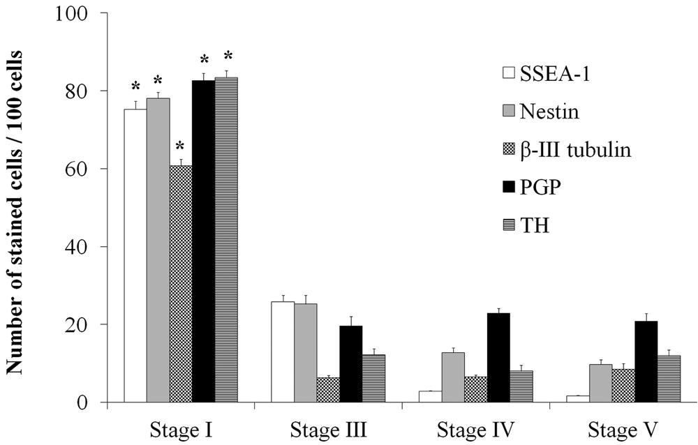
{"title":"Variation in neuronal differentiation of a newly isolated mouse embryonic stem cell line: a detailed immunocytochemistry study","authors":"Rubens Lene C. Tavares, Paloma Alvarenga Cortes, Camila Issa de Azevedo, Silvia Dantas Cangussú, Aroldo Fernando Camargos, Rosa Maria E. Arantes","doi":"10.1042/CBR20120002","DOIUrl":null,"url":null,"abstract":"<p>Neural precursor differentiation from mouse ES (embryonic stem) cells have been demonstrated using EB (embryoid body), co-culture on stromal feeder layers, and in the absence of external inducing signals. Most of available mouse ES cell original research articles have worked with only six different cell lines. Our goals were to isolate one new mouse ES lineage, and perform a detailed immunocytochemistry study during neural differentiation, making use of an EB strategy protocol following the generation of neural progenitors, glial cells and postmitotic neurons. The dynamics of differentiation of ES cell derived neuronal precursors into differentiated glia cells and neurons were followed <i>in vitro</i> and correlated to exposure to specific elements of feeder medium. Morphological aspects of generated cellular types, including its immunocytochemical expression of differentiation markers were studied. Immuno-positivity against β-III tubulin, PGP and TH (tyrosine hydroxylase) was observed from stage I. Approximately 80% of cells were positive for TH at stage I. The first glial cell type appears in stage III. TH, PGP or β-III tubulin-positive cells with neuronal typical morphology only being seen in stage III when TH-positive cells corresponded to approximately 12% of total cells. Variations among other literature findings can be explained by the choice we made to use a newly isolated ES cell line. As colonies may behave differently during neuronal differentiation, it reinforces the necessity of studying original ES cell lines.</p>","PeriodicalId":75683,"journal":{"name":"Cell biology international reports","volume":"19 1","pages":"31-35"},"PeriodicalIF":0.0000,"publicationDate":"2013-06-25","publicationTypes":"Journal Article","fieldsOfStudy":null,"isOpenAccess":false,"openAccessPdf":"https://sci-hub-pdf.com/10.1042/CBR20120002","citationCount":"2","resultStr":null,"platform":"Semanticscholar","paperid":null,"PeriodicalName":"Cell biology international reports","FirstCategoryId":"1085","ListUrlMain":"https://onlinelibrary.wiley.com/doi/10.1042/CBR20120002","RegionNum":0,"RegionCategory":null,"ArticlePicture":[],"TitleCN":null,"AbstractTextCN":null,"PMCID":null,"EPubDate":"","PubModel":"","JCR":"","JCRName":"","Score":null,"Total":0}
引用次数: 2
Abstract
Neural precursor differentiation from mouse ES (embryonic stem) cells have been demonstrated using EB (embryoid body), co-culture on stromal feeder layers, and in the absence of external inducing signals. Most of available mouse ES cell original research articles have worked with only six different cell lines. Our goals were to isolate one new mouse ES lineage, and perform a detailed immunocytochemistry study during neural differentiation, making use of an EB strategy protocol following the generation of neural progenitors, glial cells and postmitotic neurons. The dynamics of differentiation of ES cell derived neuronal precursors into differentiated glia cells and neurons were followed in vitro and correlated to exposure to specific elements of feeder medium. Morphological aspects of generated cellular types, including its immunocytochemical expression of differentiation markers were studied. Immuno-positivity against β-III tubulin, PGP and TH (tyrosine hydroxylase) was observed from stage I. Approximately 80% of cells were positive for TH at stage I. The first glial cell type appears in stage III. TH, PGP or β-III tubulin-positive cells with neuronal typical morphology only being seen in stage III when TH-positive cells corresponded to approximately 12% of total cells. Variations among other literature findings can be explained by the choice we made to use a newly isolated ES cell line. As colonies may behave differently during neuronal differentiation, it reinforces the necessity of studying original ES cell lines.


 分享
分享

 求助内容:
求助内容: 应助结果提醒方式:
 扫码关注我们
