The influence of electromyographic biofeedback therapy on knee extension following anterior cruciate ligament reconstruction: a randomized controlled trial.
Franz Christanell, Christian Hoser, Reinhard Huber, Christian Fink, Hannu Luomajoki
{"title":"The influence of electromyographic biofeedback therapy on knee extension following anterior cruciate ligament reconstruction: a randomized controlled trial.","authors":"Franz Christanell, Christian Hoser, Reinhard Huber, Christian Fink, Hannu Luomajoki","doi":"10.1186/1758-2555-4-41","DOIUrl":null,"url":null,"abstract":"<p><strong>Unlabelled: </strong></p><p><strong>Background: </strong>Loss of knee extension and a deficit in quadriceps strength are frequently found following anterior cruciate ligament (ACL) reconstruction. The aim of this study was to investigate whether the addition of Eletromyographic Biofeedback (EMG BFB) therapy for the vastus medialis muscle to the in the early phase of the standard rehabilitation programme could improve the range of knee extension and strength after ACL reconstruction more than a standard rehabilitation programme. The correlation between EMG measurement and passive knee extension was also investigated.</p><p><strong>Method: </strong>Sixteen patients, all of whom underwent endoscopic ACL reconstruction using patellar tendon autograft, were randomly assigned to two groups:• Control group (8 patients): standard rehabilitation protocol; with full weight-bearing postoperative, knee brace (0° extension, 90° flexion), electrical stimulation, aquatics and proprioceptive training.• The EMG BFB group (8 patients): EMG BFB was added to the standard rehabilitation protocol within the first postoperative week and during each session for the next 6 weeks.Each patent attended a total of 16 outpatient physiotherapy sessions following surgery. High-Heel-Distance (HHD) Test, range of motion (ROM) and integrated EMG (iEMG) for vastus medialis were measured preoperatively, and at the 1, 2, 4 and 6-week follow ups. Additionally, knee function, swelling and pain were evaluated using standardized scoring scales.</p><p><strong>Results: </strong>At 6 weeks, passive knee extension (p < 0.002) and the HHD Test were significantly (p < 0.01) better in the EMG BFB group compared to controls. Integrated EMG (vastus medialis) of the EMG BFB group also showed a significant increase after 2 (p < 0.01) and 6 (p < 0.01) weeks. At the 6-week follow up, no significant (p > 0.01) differences were found between the two groups for the assessment of knee function, swelling and pain.</p><p><strong>Conclusion: </strong>The results indicate that EMG BFB therapy, in the early phase of rehabilitation after ACL reconstruction, is useful in enhancing knee extension. Improved innervation of the vastus medialis can play a key role in the development of postoperative knee extension. EMG BFB therapy is a simple, inexpensive and valuable adjunct to conventional therapeutic modalities.</p>","PeriodicalId":88316,"journal":{"name":"Sports medicine, arthroscopy, rehabilitation, therapy & technology : SMARTT","volume":"4 1","pages":"41"},"PeriodicalIF":0.0000,"publicationDate":"2012-11-06","publicationTypes":"Journal Article","fieldsOfStudy":null,"isOpenAccess":false,"openAccessPdf":"https://sci-hub-pdf.com/10.1186/1758-2555-4-41","citationCount":"43","resultStr":null,"platform":"Semanticscholar","paperid":null,"PeriodicalName":"Sports medicine, arthroscopy, rehabilitation, therapy & technology : SMARTT","FirstCategoryId":"1085","ListUrlMain":"https://doi.org/10.1186/1758-2555-4-41","RegionNum":0,"RegionCategory":null,"ArticlePicture":[],"TitleCN":null,"AbstractTextCN":null,"PMCID":null,"EPubDate":"","PubModel":"","JCR":"","JCRName":"","Score":null,"Total":0}
引用次数: 43
Abstract
Unlabelled:
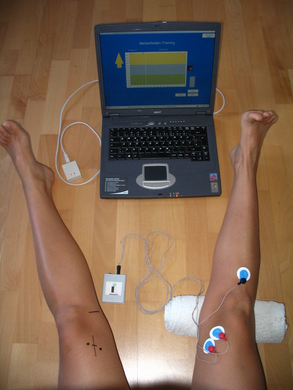
Background: Loss of knee extension and a deficit in quadriceps strength are frequently found following anterior cruciate ligament (ACL) reconstruction. The aim of this study was to investigate whether the addition of Eletromyographic Biofeedback (EMG BFB) therapy for the vastus medialis muscle to the in the early phase of the standard rehabilitation programme could improve the range of knee extension and strength after ACL reconstruction more than a standard rehabilitation programme. The correlation between EMG measurement and passive knee extension was also investigated.
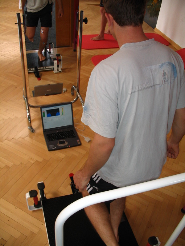
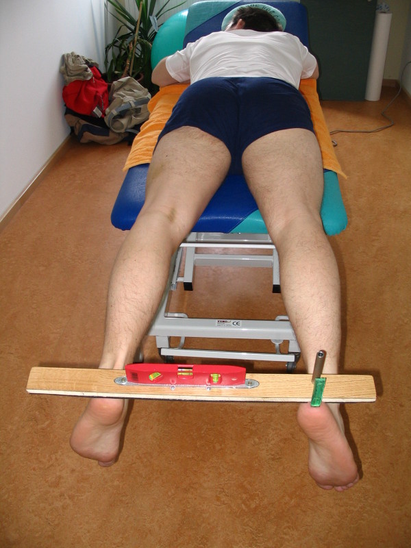
Method: Sixteen patients, all of whom underwent endoscopic ACL reconstruction using patellar tendon autograft, were randomly assigned to two groups:• Control group (8 patients): standard rehabilitation protocol; with full weight-bearing postoperative, knee brace (0° extension, 90° flexion), electrical stimulation, aquatics and proprioceptive training.• The EMG BFB group (8 patients): EMG BFB was added to the standard rehabilitation protocol within the first postoperative week and during each session for the next 6 weeks.Each patent attended a total of 16 outpatient physiotherapy sessions following surgery. High-Heel-Distance (HHD) Test, range of motion (ROM) and integrated EMG (iEMG) for vastus medialis were measured preoperatively, and at the 1, 2, 4 and 6-week follow ups. Additionally, knee function, swelling and pain were evaluated using standardized scoring scales.
Results: At 6 weeks, passive knee extension (p < 0.002) and the HHD Test were significantly (p < 0.01) better in the EMG BFB group compared to controls. Integrated EMG (vastus medialis) of the EMG BFB group also showed a significant increase after 2 (p < 0.01) and 6 (p < 0.01) weeks. At the 6-week follow up, no significant (p > 0.01) differences were found between the two groups for the assessment of knee function, swelling and pain.
Conclusion: The results indicate that EMG BFB therapy, in the early phase of rehabilitation after ACL reconstruction, is useful in enhancing knee extension. Improved innervation of the vastus medialis can play a key role in the development of postoperative knee extension. EMG BFB therapy is a simple, inexpensive and valuable adjunct to conventional therapeutic modalities.


 分享
分享

 求助内容:
求助内容: 应助结果提醒方式:
 扫码关注我们
