{"title":"Simple method for confirming tibial osteotomy during total knee arthroplasty.","authors":"Hirotaka Mutsuzaki, Kotaro Ikeda","doi":"10.1186/1758-2555-4-44","DOIUrl":null,"url":null,"abstract":"<p><strong>Unlabelled: </strong></p><p><strong>Background: </strong>Achieving precise implant alignment is crucial for producing good outcomes after total knee arthroplasty (TKA). We introduce a simple method for confirming the accuracy of tibial osteotomy during TKA.</p><p><strong>Findings: </strong>Two metallic markers were placed on the skin 20 cm apart, one on the tibial tuberosity and other on the tibial crest, points that are easily identified and palpated intraoperatively. Anteroposterior radiographs of the legs were obtained. We defined the line along the markers as the tuberosity line. The osteotomy line is perpendicular to the anatomical axis of the tibia. We then calculated the angle between these two lines and designated it the osteotomy angle. We set the osteotomy angle of the protractor, and cut the bone parallel to the osteotomy line of the protractor. Postoperatively, we analyzed the varus angle of the tibial osteotomy in 35 TKAs using the protractor. The average of the varus angle of the tibial osteotomy was 89.4° ± 1.6° (95% confidence interval of -1.0976, 0.0119). There was no significant difference from the target angle of 90° (p = 0.055). The varus angles of 90° and 90° ± 2° for the tibial osteotomy were 42.9% and 82.9%, respectively.</p><p><strong>Conclusions: </strong>We determined the accuracy of the tibial osteotomy in the coronal plane using the protractor to be satisfactory.</p>","PeriodicalId":88316,"journal":{"name":"Sports medicine, arthroscopy, rehabilitation, therapy & technology : SMARTT","volume":"4 1","pages":"44"},"PeriodicalIF":0.0000,"publicationDate":"2012-11-15","publicationTypes":"Journal Article","fieldsOfStudy":null,"isOpenAccess":false,"openAccessPdf":"https://sci-hub-pdf.com/10.1186/1758-2555-4-44","citationCount":"0","resultStr":null,"platform":"Semanticscholar","paperid":null,"PeriodicalName":"Sports medicine, arthroscopy, rehabilitation, therapy & technology : SMARTT","FirstCategoryId":"1085","ListUrlMain":"https://doi.org/10.1186/1758-2555-4-44","RegionNum":0,"RegionCategory":null,"ArticlePicture":[],"TitleCN":null,"AbstractTextCN":null,"PMCID":null,"EPubDate":"","PubModel":"","JCR":"","JCRName":"","Score":null,"Total":0}
引用次数: 0
Abstract
Unlabelled:
Background: Achieving precise implant alignment is crucial for producing good outcomes after total knee arthroplasty (TKA). We introduce a simple method for confirming the accuracy of tibial osteotomy during TKA.
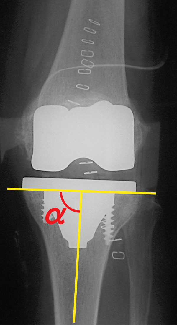
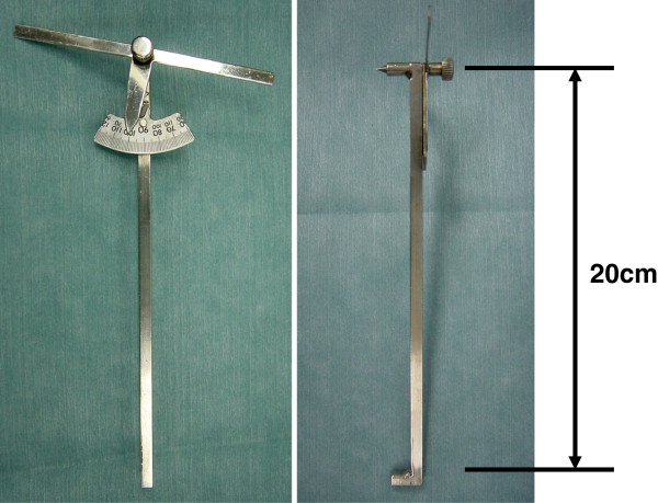
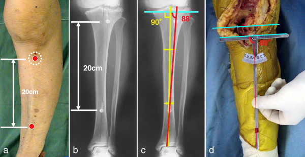
Findings: Two metallic markers were placed on the skin 20 cm apart, one on the tibial tuberosity and other on the tibial crest, points that are easily identified and palpated intraoperatively. Anteroposterior radiographs of the legs were obtained. We defined the line along the markers as the tuberosity line. The osteotomy line is perpendicular to the anatomical axis of the tibia. We then calculated the angle between these two lines and designated it the osteotomy angle. We set the osteotomy angle of the protractor, and cut the bone parallel to the osteotomy line of the protractor. Postoperatively, we analyzed the varus angle of the tibial osteotomy in 35 TKAs using the protractor. The average of the varus angle of the tibial osteotomy was 89.4° ± 1.6° (95% confidence interval of -1.0976, 0.0119). There was no significant difference from the target angle of 90° (p = 0.055). The varus angles of 90° and 90° ± 2° for the tibial osteotomy were 42.9% and 82.9%, respectively.
Conclusions: We determined the accuracy of the tibial osteotomy in the coronal plane using the protractor to be satisfactory.


 分享
分享

 求助内容:
求助内容: 应助结果提醒方式:
 扫码关注我们
