S Allen Counter, Sahar Nikkhou, Stefan Brené, Peter Damberg, Adam Sierakowiak, Tomas Klason, Cecilia Engmér Berglin, Göran Laurell
{"title":"MRI evidence of endolymphatic impermeability to the gadolinium molecule in the in vivo mouse inner ear at 9.4 tesla.","authors":"S Allen Counter, Sahar Nikkhou, Stefan Brené, Peter Damberg, Adam Sierakowiak, Tomas Klason, Cecilia Engmér Berglin, Göran Laurell","doi":"10.2174/1874440001307010027","DOIUrl":null,"url":null,"abstract":"<p><strong>Objective: </strong>Previous in vivo experimental magnetic resonance imaging (MRI) investigations of the mammalian inner ear at 4.7 Tesla have indicated that intravenously injected gadolinium (Gd) penetrates the perilymphatic labyrinth, but not the endolymphatic membranous labyrinth. In the present study, high field MRI at 9.4T was used to visualize the in vivo mouse vestibulo-cochlea system, and to determine whether the endolymphatic system is permeable to a Gd complex.</p><p><strong>Methods: </strong>A 9.4 T Varian magnet equipped with a 12 cm inner diameter gradient system with maximum gradient strength of 600 mT/m, a millipede coil (Varian design) and a Gd contrast agent were used for image acquisition in the normal C57 BL-6 mouse.</p><p><strong>Results: </strong>High-resolution 2D and 3D images of the mouse cochlea were acquired within 80 minutes following intravenous injection of Gd. Gd initially permeated the perilymphatic scala tympani and scala vestibuli, and permitted visualization of both cochlear turns from base to apex. The superior, inferior and lateral semicircular canals were subsequently visualized in 3 planes. The membranous endolymphatic labyrinth was impermeable to intravenously injected Gd, and thus showed no apparent uptake of Gd at 9.4T.</p><p><strong>Conclusion: </strong>The 9.4T field strength MRI permitted acquisition of high resolution images of anatomical and physiological features of the normal, wild type mouse perilymphatic inner ear in vivo, and provided further evidence that the endolymphatic system is impermeable to intravenously injected Gd.</p>","PeriodicalId":37431,"journal":{"name":"Open Neuroimaging Journal","volume":"7 ","pages":"27-31"},"PeriodicalIF":0.0000,"publicationDate":"2013-06-28","publicationTypes":"Journal Article","fieldsOfStudy":null,"isOpenAccess":false,"openAccessPdf":"https://ftp.ncbi.nlm.nih.gov/pub/pmc/oa_pdf/1d/6d/TONIJ-7-27.PMC3722534.pdf","citationCount":"9","resultStr":null,"platform":"Semanticscholar","paperid":null,"PeriodicalName":"Open Neuroimaging Journal","FirstCategoryId":"1085","ListUrlMain":"https://doi.org/10.2174/1874440001307010027","RegionNum":0,"RegionCategory":null,"ArticlePicture":[],"TitleCN":null,"AbstractTextCN":null,"PMCID":null,"EPubDate":"2013/1/1 0:00:00","PubModel":"Print","JCR":"Q4","JCRName":"Medicine","Score":null,"Total":0}
引用次数: 9
Abstract
Objective: Previous in vivo experimental magnetic resonance imaging (MRI) investigations of the mammalian inner ear at 4.7 Tesla have indicated that intravenously injected gadolinium (Gd) penetrates the perilymphatic labyrinth, but not the endolymphatic membranous labyrinth. In the present study, high field MRI at 9.4T was used to visualize the in vivo mouse vestibulo-cochlea system, and to determine whether the endolymphatic system is permeable to a Gd complex.
Methods: A 9.4 T Varian magnet equipped with a 12 cm inner diameter gradient system with maximum gradient strength of 600 mT/m, a millipede coil (Varian design) and a Gd contrast agent were used for image acquisition in the normal C57 BL-6 mouse.
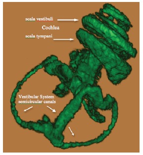
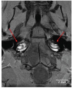
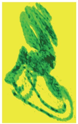
Results: High-resolution 2D and 3D images of the mouse cochlea were acquired within 80 minutes following intravenous injection of Gd. Gd initially permeated the perilymphatic scala tympani and scala vestibuli, and permitted visualization of both cochlear turns from base to apex. The superior, inferior and lateral semicircular canals were subsequently visualized in 3 planes. The membranous endolymphatic labyrinth was impermeable to intravenously injected Gd, and thus showed no apparent uptake of Gd at 9.4T.
Conclusion: The 9.4T field strength MRI permitted acquisition of high resolution images of anatomical and physiological features of the normal, wild type mouse perilymphatic inner ear in vivo, and provided further evidence that the endolymphatic system is impermeable to intravenously injected Gd.
期刊介绍:
The Open Neuroimaging Journal is an Open Access online journal, which publishes research articles, reviews/mini-reviews, and letters in all important areas of brain function, structure and organization including neuroimaging, neuroradiology, analysis methods, functional MRI acquisition and physics, brain mapping, macroscopic level of brain organization, computational modeling and analysis, structure-function and brain-behavior relationships, anatomy and physiology, psychiatric diseases and disorders of the nervous system, use of imaging to the understanding of brain pathology and brain abnormalities, cognition and aging, social neuroscience, sensorimotor processing, communication and learning.


 分享
分享
 求助内容:
求助内容: 应助结果提醒方式:
 扫码关注我们
