Emma Muñoz-Sáez, Estefanía de Munck, Paloma Maganto, Cristina Escudero, Begoña G Miguel, Rosa María Arahuetes
{"title":"In utero hepatocellular transplantation in rats.","authors":"Emma Muñoz-Sáez, Estefanía de Munck, Paloma Maganto, Cristina Escudero, Begoña G Miguel, Rosa María Arahuetes","doi":"10.1155/2013/562037","DOIUrl":null,"url":null,"abstract":"<p><p>This work represents a step forward in the experimental design of an in utero hepatocellular transplantation model in rats. We focused on the enrichment optimization of isolated fetal hepatocytes suspension, arranging the surgery methodology of in utero transplantation, monitoring the biodistribution of the transplanted hepatocytes, and assessing the success of the transplants. Rat fetuses have been transplanted at the 17th embryonic day (ED17) with fetal hepatocytes isolated from rats at the end of pregnancy (ED21). We assessed possible differences between lymphocyte population, CD4 positive, CD8 positive, double-positive T-cells, and anti-inflammatory cytokines interleukins 4 and 10 (IL4 and IL10) as well. Cellular viability reached the rates of 90-95%. Transplanted groups had a limited success. Transplanted hepatocytes were not able to pass through the hematoplacental barrier. The hepatocytes injected were primarily located in the liver. There was an upward trend in the whole amount of T CD4 and T CD8 cells. There was an increased IL4 in the transplanted groups observed in the pregnant rats. The possibility to induce tolerance in fetuses with a hepatocyte transplant in utero could be a key point to avoid the immunosuppression treatments which must be undergone by transplanted patients.</p>","PeriodicalId":55254,"journal":{"name":"Clinical & Developmental Immunology","volume":"2013 ","pages":"562037"},"PeriodicalIF":0.0000,"publicationDate":"2013-01-01","publicationTypes":"Journal Article","fieldsOfStudy":null,"isOpenAccess":false,"openAccessPdf":"https://sci-hub-pdf.com/10.1155/2013/562037","citationCount":"3","resultStr":null,"platform":"Semanticscholar","paperid":null,"PeriodicalName":"Clinical & Developmental Immunology","FirstCategoryId":"1085","ListUrlMain":"https://doi.org/10.1155/2013/562037","RegionNum":0,"RegionCategory":null,"ArticlePicture":[],"TitleCN":null,"AbstractTextCN":null,"PMCID":null,"EPubDate":"2013/8/24 0:00:00","PubModel":"Epub","JCR":"","JCRName":"","Score":null,"Total":0}
引用次数: 3
Abstract
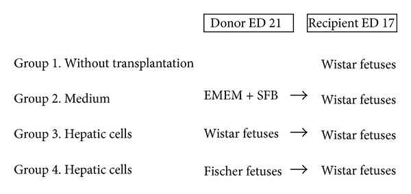
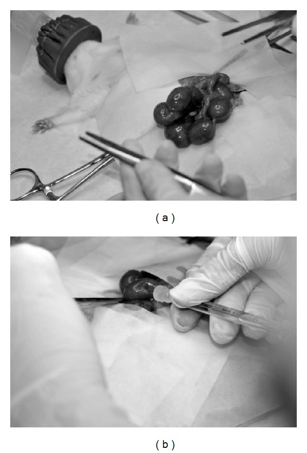
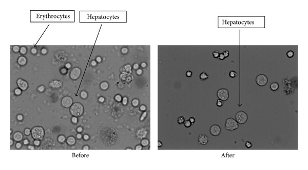
This work represents a step forward in the experimental design of an in utero hepatocellular transplantation model in rats. We focused on the enrichment optimization of isolated fetal hepatocytes suspension, arranging the surgery methodology of in utero transplantation, monitoring the biodistribution of the transplanted hepatocytes, and assessing the success of the transplants. Rat fetuses have been transplanted at the 17th embryonic day (ED17) with fetal hepatocytes isolated from rats at the end of pregnancy (ED21). We assessed possible differences between lymphocyte population, CD4 positive, CD8 positive, double-positive T-cells, and anti-inflammatory cytokines interleukins 4 and 10 (IL4 and IL10) as well. Cellular viability reached the rates of 90-95%. Transplanted groups had a limited success. Transplanted hepatocytes were not able to pass through the hematoplacental barrier. The hepatocytes injected were primarily located in the liver. There was an upward trend in the whole amount of T CD4 and T CD8 cells. There was an increased IL4 in the transplanted groups observed in the pregnant rats. The possibility to induce tolerance in fetuses with a hepatocyte transplant in utero could be a key point to avoid the immunosuppression treatments which must be undergone by transplanted patients.


 分享
分享
 求助内容:
求助内容: 应助结果提醒方式:
 扫码关注我们
