{"title":"Cystic benign phyllodes tumor in the inguinal region.","authors":"Jai Hyang Go","doi":"10.4132/KoreanJPathol.2013.47.6.583","DOIUrl":null,"url":null,"abstract":"<p><p>The present lesion was the first reported case of a benign intracystic phyllodes tumor in the inguinal region. We report the case of a 51-year-old female patient who presented with an inguinal mass. A clinical diagnosis of malignant lymphoma was considered in this case. The resected tumor was well-circumscribed and showed numerous papillary nodular protrusions into a central cystic cavity (3.5×2.5 cm). The microscopic findings showed hyperplastic epithelium-lined cysts with leaf-like intraluminal epithelium-lined bland stromal projections. The epithelial cell linings were strongly positive for estrogen and progesterone receptors. </p>","PeriodicalId":49936,"journal":{"name":"Korean Journal of Pathology","volume":"47 6","pages":"583-6"},"PeriodicalIF":0.0000,"publicationDate":"2013-12-01","publicationTypes":"Journal Article","fieldsOfStudy":null,"isOpenAccess":false,"openAccessPdf":"https://sci-hub-pdf.com/10.4132/KoreanJPathol.2013.47.6.583","citationCount":"2","resultStr":null,"platform":"Semanticscholar","paperid":null,"PeriodicalName":"Korean Journal of Pathology","FirstCategoryId":"1085","ListUrlMain":"https://doi.org/10.4132/KoreanJPathol.2013.47.6.583","RegionNum":0,"RegionCategory":null,"ArticlePicture":[],"TitleCN":null,"AbstractTextCN":null,"PMCID":null,"EPubDate":"2013/12/24 0:00:00","PubModel":"Epub","JCR":"","JCRName":"","Score":null,"Total":0}
引用次数: 2
Abstract
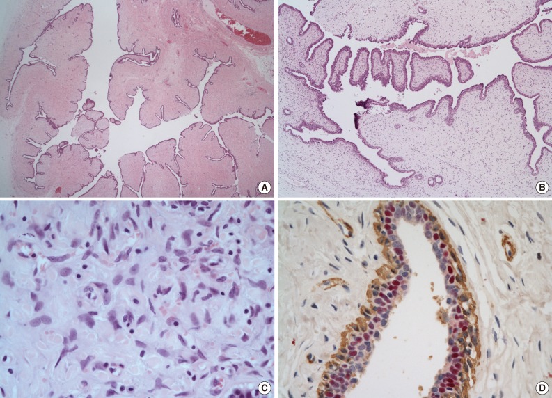
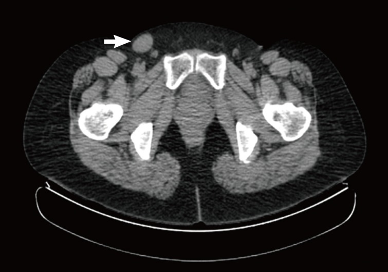
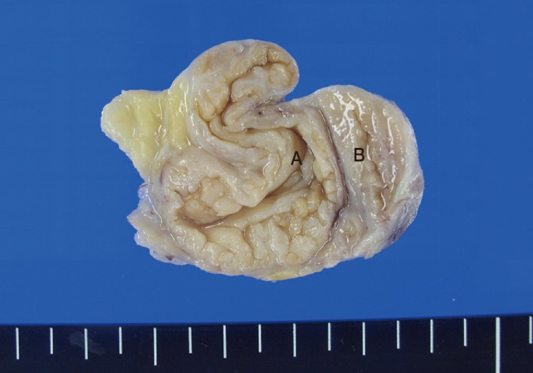
The present lesion was the first reported case of a benign intracystic phyllodes tumor in the inguinal region. We report the case of a 51-year-old female patient who presented with an inguinal mass. A clinical diagnosis of malignant lymphoma was considered in this case. The resected tumor was well-circumscribed and showed numerous papillary nodular protrusions into a central cystic cavity (3.5×2.5 cm). The microscopic findings showed hyperplastic epithelium-lined cysts with leaf-like intraluminal epithelium-lined bland stromal projections. The epithelial cell linings were strongly positive for estrogen and progesterone receptors.


 分享
分享
 求助内容:
求助内容: 应助结果提醒方式:
 扫码关注我们
