Brad A Culotta, Donald A Deinlein, Steven M Theiss, Jack E Lemons
{"title":"An extension-distraction injury of the thoracic spine with traumatic partial correction of thoracic kyphosis.","authors":"Brad A Culotta, Donald A Deinlein, Steven M Theiss, Jack E Lemons","doi":"10.1055/s-0033-1347132","DOIUrl":null,"url":null,"abstract":"<p><p>Study Design The study is a case report. Objective The authors aim to report an unusual injury pattern in a patient previously treated for thoracic kyphoscoliosis. Methods A postoperative (computed tomography) CT of a healthy 24-year-old man who underwent posterior instrumentation and fusion for a kyphoscoliosis deformity was compared with a CT performed after a motor vehicle accident (MVA) 1 year later, which resulted in an extension-distraction injury of T8 with no neurologic deficit. Cobb angles of the thoracic sagittal images of both CTs were measured using a digital measuring device and the values were recorded. Results Initial postoperative sagittal CT images demonstrate a 67-degree residual thoracic kyphosis compared with the post-MVA sagittal CT images, which reveal a 54-degree thoracic kyphosis, a 13-degree improvement in sagittal alignment. Conclusion It is unusual for a patient with long posterior instrumentation of the spine to sustain a spinal fracture without breakage of the rods, which were 6-mm nickel-titanium alloy with two crosslinks. Although sustaining plastic deformation, the rods maintained their integrity to the degree that the patient required no subsequent treatment to his spine at 12 months follow-up. It is rare to sustain a vertebral fracture without implant failure, which occurred in this case. </p>","PeriodicalId":89675,"journal":{"name":"Evidence-based spine-care journal","volume":"4 2","pages":"126-31"},"PeriodicalIF":0.0000,"publicationDate":"2013-10-01","publicationTypes":"Journal Article","fieldsOfStudy":null,"isOpenAccess":false,"openAccessPdf":"https://sci-hub-pdf.com/10.1055/s-0033-1347132","citationCount":"0","resultStr":null,"platform":"Semanticscholar","paperid":null,"PeriodicalName":"Evidence-based spine-care journal","FirstCategoryId":"1085","ListUrlMain":"https://doi.org/10.1055/s-0033-1347132","RegionNum":0,"RegionCategory":null,"ArticlePicture":[],"TitleCN":null,"AbstractTextCN":null,"PMCID":null,"EPubDate":"2013/6/18 0:00:00","PubModel":"Epub","JCR":"","JCRName":"","Score":null,"Total":0}
引用次数: 0
Abstract
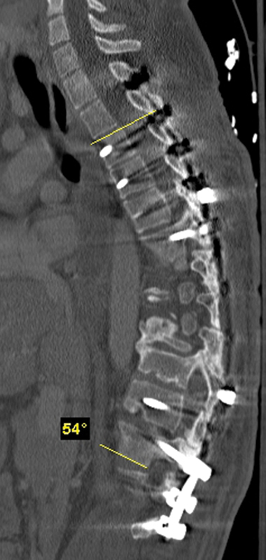
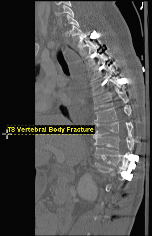
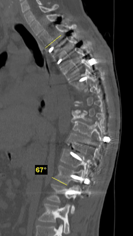
Study Design The study is a case report. Objective The authors aim to report an unusual injury pattern in a patient previously treated for thoracic kyphoscoliosis. Methods A postoperative (computed tomography) CT of a healthy 24-year-old man who underwent posterior instrumentation and fusion for a kyphoscoliosis deformity was compared with a CT performed after a motor vehicle accident (MVA) 1 year later, which resulted in an extension-distraction injury of T8 with no neurologic deficit. Cobb angles of the thoracic sagittal images of both CTs were measured using a digital measuring device and the values were recorded. Results Initial postoperative sagittal CT images demonstrate a 67-degree residual thoracic kyphosis compared with the post-MVA sagittal CT images, which reveal a 54-degree thoracic kyphosis, a 13-degree improvement in sagittal alignment. Conclusion It is unusual for a patient with long posterior instrumentation of the spine to sustain a spinal fracture without breakage of the rods, which were 6-mm nickel-titanium alloy with two crosslinks. Although sustaining plastic deformation, the rods maintained their integrity to the degree that the patient required no subsequent treatment to his spine at 12 months follow-up. It is rare to sustain a vertebral fracture without implant failure, which occurred in this case.


 分享
分享

 求助内容:
求助内容: 应助结果提醒方式:
 扫码关注我们
