Joseph S Cheng, Christopher B Carr, Cyrus Wong, Adrija Sharma, Mohamed R Mahfouz, Richard D Komistek
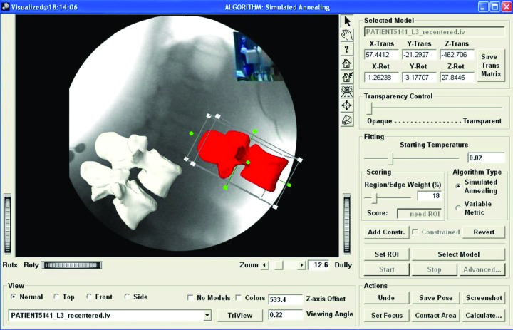
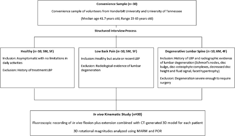
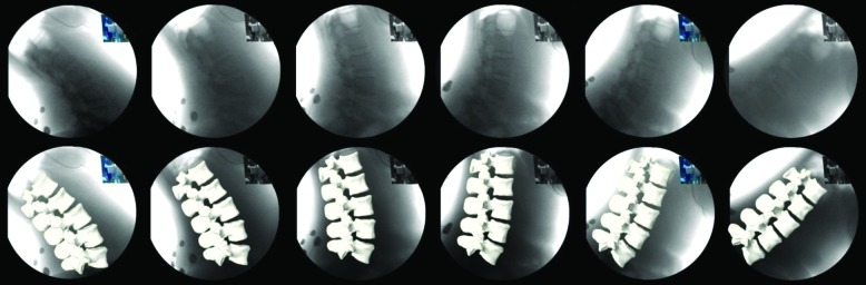
{"title":"Altered spinal motion in low back pain associated with lumbar strain and spondylosis.","authors":"Joseph S Cheng, Christopher B Carr, Cyrus Wong, Adrija Sharma, Mohamed R Mahfouz, Richard D Komistek","doi":"10.1055/s-0033-1341640","DOIUrl":null,"url":null,"abstract":"<p><p>Study Design We present a patient-specific computer model created to translate two-dimensional (2D) fluoroscopic motion data into three-dimensional (3D) in vivo biomechanical motion data. Objective The aim of this study is to determine the in vivo biomechanical differences in patients with and without acute low back pain. Current dynamic imaging of the lumbar spine consists of flexion-extension static radiographs, which lack sensitivity to out-of-plane motion and provide incomplete information on the overall spinal motion. Using a novel technique, in-plane and coupled out-of-plane rotational motions are quantified in the lumbar spine. Methods A total of 30 participants-10 healthy asymptomatic subjects, 10 patients with low back pain without spondylosis radiologically, and 10 patients with low back pain with radiological spondylosis-underwent dynamic fluoroscopy with a 3D-to-2D image registration technique to create a 3D, patient-specific bone model to analyze in vivo kinematics using the maximal absolute rotational magnitude and the path of rotation. Results Average overall in-plane rotations (L1-L5) in patients with low back pain were less than those asymptomatic, with the dominant loss of motion during extension. Those with low back pain also had significantly greater out-of-plane rotations, with 5.5 degrees (without spondylosis) and 7.1 degrees (with spondylosis) more out-of-plane rotational motion per level compared with asymptomatic subjects. Conclusions Subjects with low back pain exhibited greater out-of-plane intersegmental motion in their lumbar spine than healthy asymptomatic subjects. Conventional flexion-extension radiographs are inadequate for evaluating motion patterns of lumbar strain, and assessment of 3D in vivo spinal motion may elucidate the association of abnormal vertebral motions and clinically significant low back pain. </p>","PeriodicalId":89675,"journal":{"name":"Evidence-based spine-care journal","volume":"4 1","pages":"6-12"},"PeriodicalIF":0.0000,"publicationDate":"2013-04-01","publicationTypes":"Journal Article","fieldsOfStudy":null,"isOpenAccess":false,"openAccessPdf":"https://sci-hub-pdf.com/10.1055/s-0033-1341640","citationCount":"11","resultStr":null,"platform":"Semanticscholar","paperid":null,"PeriodicalName":"Evidence-based spine-care journal","FirstCategoryId":"1085","ListUrlMain":"https://doi.org/10.1055/s-0033-1341640","RegionNum":0,"RegionCategory":null,"ArticlePicture":[],"TitleCN":null,"AbstractTextCN":null,"PMCID":null,"EPubDate":"","PubModel":"","JCR":"","JCRName":"","Score":null,"Total":0}
引用次数: 11
Abstract
Study Design We present a patient-specific computer model created to translate two-dimensional (2D) fluoroscopic motion data into three-dimensional (3D) in vivo biomechanical motion data. Objective The aim of this study is to determine the in vivo biomechanical differences in patients with and without acute low back pain. Current dynamic imaging of the lumbar spine consists of flexion-extension static radiographs, which lack sensitivity to out-of-plane motion and provide incomplete information on the overall spinal motion. Using a novel technique, in-plane and coupled out-of-plane rotational motions are quantified in the lumbar spine. Methods A total of 30 participants-10 healthy asymptomatic subjects, 10 patients with low back pain without spondylosis radiologically, and 10 patients with low back pain with radiological spondylosis-underwent dynamic fluoroscopy with a 3D-to-2D image registration technique to create a 3D, patient-specific bone model to analyze in vivo kinematics using the maximal absolute rotational magnitude and the path of rotation. Results Average overall in-plane rotations (L1-L5) in patients with low back pain were less than those asymptomatic, with the dominant loss of motion during extension. Those with low back pain also had significantly greater out-of-plane rotations, with 5.5 degrees (without spondylosis) and 7.1 degrees (with spondylosis) more out-of-plane rotational motion per level compared with asymptomatic subjects. Conclusions Subjects with low back pain exhibited greater out-of-plane intersegmental motion in their lumbar spine than healthy asymptomatic subjects. Conventional flexion-extension radiographs are inadequate for evaluating motion patterns of lumbar strain, and assessment of 3D in vivo spinal motion may elucidate the association of abnormal vertebral motions and clinically significant low back pain.


 分享
分享

 求助内容:
求助内容: 应助结果提醒方式:
 扫码关注我们
