Yang Liu, Shikhar Uttam, Sergey Alexandrov, Rajan K Bista
{"title":"Investigation of nanoscale structural alterations of cell nucleus as an early sign of cancer.","authors":"Yang Liu, Shikhar Uttam, Sergey Alexandrov, Rajan K Bista","doi":"10.1186/2046-1682-7-1","DOIUrl":null,"url":null,"abstract":"<p><strong>Background: </strong>The cell and tissue structural properties assessed with a conventional bright-field light microscope play a key role in cancer diagnosis, but they sometimes have limited accuracy in detecting early-stage cancers or predicting future risk of cancer progression for individual patients (i.e., prognosis) if no frank cancer is found. The recent development in optical microscopy techniques now permit the nanoscale structural imaging and quantitative structural analysis of tissue and cells, which offers a new opportunity to investigate the structural properties of cell and tissue below 200 - 250 nm as an early sign of carcinogenesis, prior to the presence of microscale morphological abnormalities. Identification of nanoscale structural signatures is significant for earlier and more accurate cancer detection and prognosis.</p><p><strong>Results: </strong>Our group has recently developed two simple spectral-domain optical microscopy techniques for assessing 3D nanoscale structural alterations - spectral-encoding of spatial frequency microscopy and spatial-domain low-coherence quantitative phase microscopy. These two techniques use the scattered light from biological cells and tissue and share a common experimental approach of assessing the Fourier space by various wavelengths to quantify the 3D structural information of the scattering object at the nanoscale sensitivity with a simple reflectance-mode light microscopy setup without the need for high-NA optics. This review paper discusses the physical principles and validation of these two techniques to interrogate nanoscale structural properties, as well as the use of these methods to probe nanoscale nuclear architectural alterations during carcinogenesis in cancer cell lines and well-annotated human tissue during carcinogenesis.</p><p><strong>Conclusions: </strong>The analysis of nanoscale structural characteristics has shown promise in detecting cancer before the microscopically visible changes become evident and proof-of-concept studies have shown its feasibility as an earlier or more sensitive marker for cancer detection or diagnosis. Further biophysical investigation of specific 3D nanoscale structural characteristics in carcinogenesis, especially with well-annotated human cells and tissue, is much needed in cancer research.</p>","PeriodicalId":9045,"journal":{"name":"BMC Biophysics","volume":"7 1","pages":"1"},"PeriodicalIF":0.0000,"publicationDate":"2014-02-10","publicationTypes":"Journal Article","fieldsOfStudy":null,"isOpenAccess":false,"openAccessPdf":"https://sci-hub-pdf.com/10.1186/2046-1682-7-1","citationCount":"18","resultStr":null,"platform":"Semanticscholar","paperid":null,"PeriodicalName":"BMC Biophysics","FirstCategoryId":"1085","ListUrlMain":"https://doi.org/10.1186/2046-1682-7-1","RegionNum":0,"RegionCategory":null,"ArticlePicture":[],"TitleCN":null,"AbstractTextCN":null,"PMCID":null,"EPubDate":"","PubModel":"","JCR":"Q1","JCRName":"Biochemistry, Genetics and Molecular Biology","Score":null,"Total":0}
引用次数: 18
Abstract
Background: The cell and tissue structural properties assessed with a conventional bright-field light microscope play a key role in cancer diagnosis, but they sometimes have limited accuracy in detecting early-stage cancers or predicting future risk of cancer progression for individual patients (i.e., prognosis) if no frank cancer is found. The recent development in optical microscopy techniques now permit the nanoscale structural imaging and quantitative structural analysis of tissue and cells, which offers a new opportunity to investigate the structural properties of cell and tissue below 200 - 250 nm as an early sign of carcinogenesis, prior to the presence of microscale morphological abnormalities. Identification of nanoscale structural signatures is significant for earlier and more accurate cancer detection and prognosis.
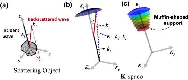
Results: Our group has recently developed two simple spectral-domain optical microscopy techniques for assessing 3D nanoscale structural alterations - spectral-encoding of spatial frequency microscopy and spatial-domain low-coherence quantitative phase microscopy. These two techniques use the scattered light from biological cells and tissue and share a common experimental approach of assessing the Fourier space by various wavelengths to quantify the 3D structural information of the scattering object at the nanoscale sensitivity with a simple reflectance-mode light microscopy setup without the need for high-NA optics. This review paper discusses the physical principles and validation of these two techniques to interrogate nanoscale structural properties, as well as the use of these methods to probe nanoscale nuclear architectural alterations during carcinogenesis in cancer cell lines and well-annotated human tissue during carcinogenesis.
Conclusions: The analysis of nanoscale structural characteristics has shown promise in detecting cancer before the microscopically visible changes become evident and proof-of-concept studies have shown its feasibility as an earlier or more sensitive marker for cancer detection or diagnosis. Further biophysical investigation of specific 3D nanoscale structural characteristics in carcinogenesis, especially with well-annotated human cells and tissue, is much needed in cancer research.


 分享
分享
 求助内容:
求助内容: 应助结果提醒方式:
 扫码关注我们
