{"title":"The role of elastosonography in the differentiation of parotid gland lesions: report of three cases and review of the literature.","authors":"Idil Gunes Tatar, Onur Ergun, Aydin Kurt, Mustafa Sahin, Baki Hekimoğlu","doi":"10.12659/PJR.891019","DOIUrl":null,"url":null,"abstract":"<p><strong>Background: </strong>The parotid gland is the mostly affected site among major salivary gland tumors in up to 85% of cases. Preoperative knowledge of the tumour nature is crucial since it influences the surgical procedure and patient's morbidity, especially the risk of facial nerve palsy. Ultrasonography is commonly used as the first line imaging modality for the salivary gland lesions. A pitfall is that the histologic pleomorphism often reflects an imaging pleomorphism.</p><p><strong>Case report: </strong>HEREIN WE AIMED TO PRESENT THE ROLE OF ELASTOSONOGRAPHY IN THREE PAROTID LESIONS: a case of benign pleomorphic adenoma, a Wharthin's tumour and a malignant parotid tumour.</p><p><strong>Conclusions: </strong>Our findings show that malignant parotid lesion was the stiffest lesion according to elastosonography. Wharthin's tumour demonstrated soft elastosonographic features. The pleomorphic adenoma was also interpreted as stiff by elastosonography suggesting that the elastosonographic features of pleomorphic adenoma may resemble those of malignant lesions limiting the utility of the technique.</p>","PeriodicalId":47128,"journal":{"name":"Polish Journal of Radiology","volume":"79 ","pages":"398-401"},"PeriodicalIF":1.6000,"publicationDate":"2014-11-06","publicationTypes":"Journal Article","fieldsOfStudy":null,"isOpenAccess":false,"openAccessPdf":"https://www.ncbi.nlm.nih.gov/pmc/articles/PMC4226314/pdf/","citationCount":"7","resultStr":null,"platform":"Semanticscholar","paperid":null,"PeriodicalName":"Polish Journal of Radiology","FirstCategoryId":"1085","ListUrlMain":"https://doi.org/10.12659/PJR.891019","RegionNum":0,"RegionCategory":null,"ArticlePicture":[],"TitleCN":null,"AbstractTextCN":null,"PMCID":null,"EPubDate":"2014/1/1 0:00:00","PubModel":"eCollection","JCR":"Q4","JCRName":"RADIOLOGY, NUCLEAR MEDICINE & MEDICAL IMAGING","Score":null,"Total":0}
引用次数: 7
Abstract
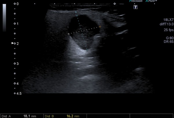
Background: The parotid gland is the mostly affected site among major salivary gland tumors in up to 85% of cases. Preoperative knowledge of the tumour nature is crucial since it influences the surgical procedure and patient's morbidity, especially the risk of facial nerve palsy. Ultrasonography is commonly used as the first line imaging modality for the salivary gland lesions. A pitfall is that the histologic pleomorphism often reflects an imaging pleomorphism.
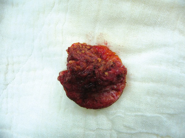
Case report: HEREIN WE AIMED TO PRESENT THE ROLE OF ELASTOSONOGRAPHY IN THREE PAROTID LESIONS: a case of benign pleomorphic adenoma, a Wharthin's tumour and a malignant parotid tumour.
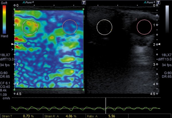
Conclusions: Our findings show that malignant parotid lesion was the stiffest lesion according to elastosonography. Wharthin's tumour demonstrated soft elastosonographic features. The pleomorphic adenoma was also interpreted as stiff by elastosonography suggesting that the elastosonographic features of pleomorphic adenoma may resemble those of malignant lesions limiting the utility of the technique.


 分享
分享
 求助内容:
求助内容: 应助结果提醒方式:
 扫码关注我们
