{"title":"Value of Optic Nerve Sheath Diameter in Diagnosis and Follow Up of Patients with Disturbed Conscious Level.","authors":"Osama Mahmoud Momtaz, Omar M Said, Amany Mahmoud Mohamed, Tamer Sayed Abdel Mawla","doi":"10.2147/EB.S369813","DOIUrl":null,"url":null,"abstract":"<p><strong>Background: </strong>Ultrasonographic measurement of optic nerve sheath diameter is a simple, non-invasive, and reliable method of detecting elevated intracranial pressure (ICP) in critical patients. Optic nerve sheath communicates with the dura mater covering the brain and contains cerebrospinal fluid, allowing pressure transmission from the cranium. Therefore, changes in cerebrospinal fluid (CSF) pressure have been shown to produce changes in ONSD.</p><p><strong>Objective: </strong>This study aimed to assess the accuracy of optic nerve sheath diameter (ONSD) in diagnosis and follow-up patients with disturbed conscious levels compared with CT brain and fundus examination.</p><p><strong>Patients and methods: </strong>One hundred forty-one participants were included in the study, classified into 76 cases admitted with disturbed conscious levels due to elevated ICP and 65 controls. All patients were subjected to CT brain and optic nerve US and fundus examination at the time of admission and follow-up after 48 h after proper management.</p><p><strong>Results: </strong>The current study showed that ONSD is significant in predicting elevated ICP at the cut-off point of average ONSD of 5.19 mm with 97% sensitivity and 98% specificity, and the area under the curve (AUC) was 0.996. The present study revealed a significant inverse correlation between ONSD and GCS in patients with increased ICP.</p><p><strong>Conclusion: </strong>Ultrasonic measurement of ONSD is a promising technique in diagnosing and following patients with disturbed conscious levels.</p>","PeriodicalId":51844,"journal":{"name":"Eye and Brain","volume":" ","pages":"115-126"},"PeriodicalIF":2.4000,"publicationDate":"2022-09-27","publicationTypes":"Journal Article","fieldsOfStudy":null,"isOpenAccess":false,"openAccessPdf":"https://ftp.ncbi.nlm.nih.gov/pub/pmc/oa_pdf/5b/4a/eb-14-115.PMC9526430.pdf","citationCount":"0","resultStr":null,"platform":"Semanticscholar","paperid":null,"PeriodicalName":"Eye and Brain","FirstCategoryId":"1085","ListUrlMain":"https://doi.org/10.2147/EB.S369813","RegionNum":0,"RegionCategory":null,"ArticlePicture":[],"TitleCN":null,"AbstractTextCN":null,"PMCID":null,"EPubDate":"2022/1/1 0:00:00","PubModel":"eCollection","JCR":"Q1","JCRName":"OPHTHALMOLOGY","Score":null,"Total":0}
引用次数: 0
Abstract
Background: Ultrasonographic measurement of optic nerve sheath diameter is a simple, non-invasive, and reliable method of detecting elevated intracranial pressure (ICP) in critical patients. Optic nerve sheath communicates with the dura mater covering the brain and contains cerebrospinal fluid, allowing pressure transmission from the cranium. Therefore, changes in cerebrospinal fluid (CSF) pressure have been shown to produce changes in ONSD.
Objective: This study aimed to assess the accuracy of optic nerve sheath diameter (ONSD) in diagnosis and follow-up patients with disturbed conscious levels compared with CT brain and fundus examination.
Patients and methods: One hundred forty-one participants were included in the study, classified into 76 cases admitted with disturbed conscious levels due to elevated ICP and 65 controls. All patients were subjected to CT brain and optic nerve US and fundus examination at the time of admission and follow-up after 48 h after proper management.
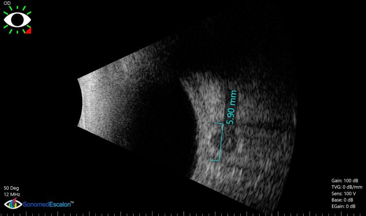
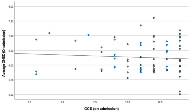
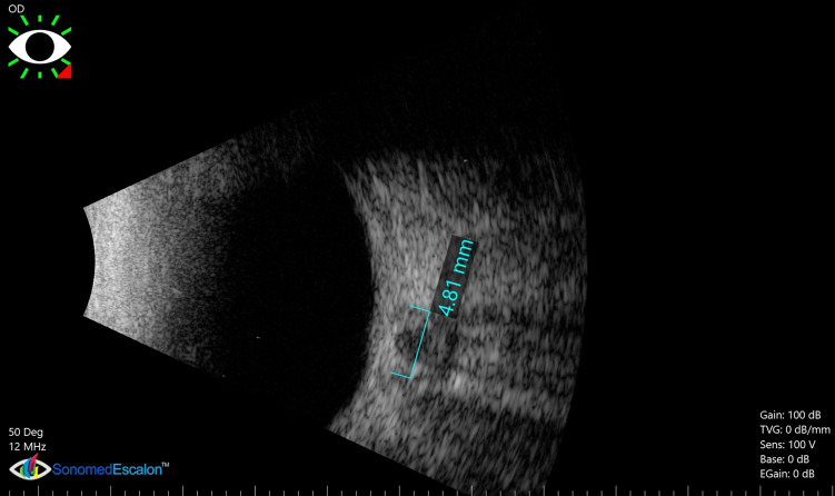
Results: The current study showed that ONSD is significant in predicting elevated ICP at the cut-off point of average ONSD of 5.19 mm with 97% sensitivity and 98% specificity, and the area under the curve (AUC) was 0.996. The present study revealed a significant inverse correlation between ONSD and GCS in patients with increased ICP.
Conclusion: Ultrasonic measurement of ONSD is a promising technique in diagnosing and following patients with disturbed conscious levels.
期刊介绍:
Eye and Brain is an international, peer-reviewed, open access journal focusing on basic research, clinical findings, and expert reviews in the field of visual science and neuro-ophthalmology. The journal’s unique focus is the link between two well-known visual centres, the eye and the brain, with an emphasis on the importance of such connections. All aspects of clinical and especially basic research on the visual system are addressed within the journal as well as significant future directions in vision research and therapeutic measures. This unique journal focuses on neurological aspects of vision – both physiological and pathological. The scope of the journal spans from the cornea to the associational visual cortex and all the visual centers in between. Topics range from basic biological mechanisms to therapeutic treatment, from simple organisms to humans, and utilizing techniques from molecular biology to behavior. The journal especially welcomes primary research articles or review papers that make the connection between the eye and the brain. Specific areas covered in the journal include: Physiology and pathophysiology of visual centers, Eye movement disorders and strabismus, Cellular, biochemical, and molecular features of the visual system, Structural and functional organization of the eye and of the visual cortex, Metabolic demands of the visual system, Diseases and disorders with neuro-ophthalmic manifestations, Clinical and experimental neuro-ophthalmology and visual system pathologies, Epidemiological studies.


 分享
分享
 求助内容:
求助内容: 应助结果提醒方式:
 扫码关注我们
