{"title":"PET-MRI in idiopathic inflammatory myositis: a comparative study of clinical and immunological markers with imaging findings.","authors":"Manu Santhappan Girija, Ravindu Tiwari, Seena Vengalil, Saraswati Nashi, Veeramani Preethish-Kumar, Kiran Polavarapu, Karthik Kulanthaivelu, Arpana Arbind, Mainak Bardhan, Akshata Huddar, Gopikrishnan Unnikrishnan, Valasani Ravi Kiran, Tanushree Chawla, Bevinahalli Nandeesh, Chandana Nagaraj, Atchayaram Nalini","doi":"10.1186/s42466-022-00213-9","DOIUrl":null,"url":null,"abstract":"<p><strong>Background: </strong>We sought to determine the utility of PET-MRI in diagnosing Idiopathic Inflammatory Myositis (IIM), and look for association between FDG uptake and clinical, pathological and laboratory parameters.</p><p><strong>Methods: </strong>A retrospective, observational study was conducted on IIM patients having positive serum autoantibodies and who underwent PET-MRI (3-Tesla SIEMENS Biograph MR scanner) between 2017 and 2021. Thirty patients who underwent PET-MRI to detect systemic metastasis without muscle involvement formed the control group.</p><p><strong>Results: </strong>In the IIM cohort, female: male sex ratio was 1.73, mean age at diagnosis was 40.33 years, and the mean duration of illness was 7 months. 33.33% of patients had severe limb weakness. Mi2B (43.33%), Mi2A (43.33%), PL-7(10%), PL-12(6.67%), SRP (16.67%), Tif1gamma (3.33%), NxP2 (3.33%), Ro-52(40%), PM-Scl, U1-RNP, ANA (26.67%) were the serum autoantibodies identified. Using SUV max Ratio to quantify FDG uptake, PET-MRI showed a sensitivity of 100% with 93.3% specificity in diagnosing IIM.FDG uptake was maximum in proximal lower limb region followed by proximal upper limb. Multivariate regression analysis showed that the severity of muscle weakness, serum Mi2B antibody positivity and serum creatinine kinase levels had a significant positive correlation with FDG uptake (value of 0.005, 0.043, 0.042, respectively for whole-body FDG uptake). FDG uptake also showed good correlation with histopathological features and muscle MRI, but there was no significant association with treatment response. Three female patients in our cohort had primary malignancy involving the breast, uterus, and cervix.</p><p><strong>Conclusions: </strong>PET-MRI is a promising diagnostic modality for IIM. PET-MRI reflects the severity of muscle inflammation, showing good association with various clinical/laboratory parameters, histopathology, and muscle MRI. Parameters associated with severe muscle inflammation in PET-MRI-clinical severity of muscle weakness, Mi2B positivity, and serum creatine kinase levels-may be used as clinical/laboratory markers of disease severity in IIM. PET-MRI has the added advantage of detection of systemic malignancy.</p>","PeriodicalId":19169,"journal":{"name":"Neurological Research and Practice","volume":" ","pages":"49"},"PeriodicalIF":0.0000,"publicationDate":"2022-10-10","publicationTypes":"Journal Article","fieldsOfStudy":null,"isOpenAccess":false,"openAccessPdf":"https://www.ncbi.nlm.nih.gov/pmc/articles/PMC9549636/pdf/","citationCount":"3","resultStr":null,"platform":"Semanticscholar","paperid":null,"PeriodicalName":"Neurological Research and Practice","FirstCategoryId":"1085","ListUrlMain":"https://doi.org/10.1186/s42466-022-00213-9","RegionNum":0,"RegionCategory":null,"ArticlePicture":[],"TitleCN":null,"AbstractTextCN":null,"PMCID":null,"EPubDate":"","PubModel":"","JCR":"","JCRName":"","Score":null,"Total":0}
引用次数: 3
Abstract
Background: We sought to determine the utility of PET-MRI in diagnosing Idiopathic Inflammatory Myositis (IIM), and look for association between FDG uptake and clinical, pathological and laboratory parameters.
Methods: A retrospective, observational study was conducted on IIM patients having positive serum autoantibodies and who underwent PET-MRI (3-Tesla SIEMENS Biograph MR scanner) between 2017 and 2021. Thirty patients who underwent PET-MRI to detect systemic metastasis without muscle involvement formed the control group.
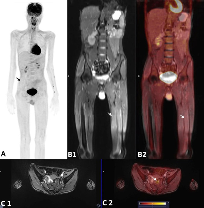
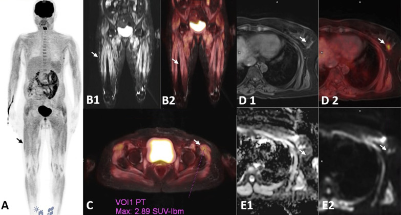
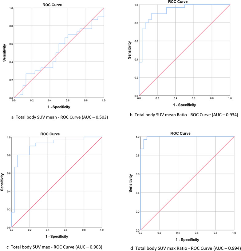
Results: In the IIM cohort, female: male sex ratio was 1.73, mean age at diagnosis was 40.33 years, and the mean duration of illness was 7 months. 33.33% of patients had severe limb weakness. Mi2B (43.33%), Mi2A (43.33%), PL-7(10%), PL-12(6.67%), SRP (16.67%), Tif1gamma (3.33%), NxP2 (3.33%), Ro-52(40%), PM-Scl, U1-RNP, ANA (26.67%) were the serum autoantibodies identified. Using SUV max Ratio to quantify FDG uptake, PET-MRI showed a sensitivity of 100% with 93.3% specificity in diagnosing IIM.FDG uptake was maximum in proximal lower limb region followed by proximal upper limb. Multivariate regression analysis showed that the severity of muscle weakness, serum Mi2B antibody positivity and serum creatinine kinase levels had a significant positive correlation with FDG uptake (value of 0.005, 0.043, 0.042, respectively for whole-body FDG uptake). FDG uptake also showed good correlation with histopathological features and muscle MRI, but there was no significant association with treatment response. Three female patients in our cohort had primary malignancy involving the breast, uterus, and cervix.
Conclusions: PET-MRI is a promising diagnostic modality for IIM. PET-MRI reflects the severity of muscle inflammation, showing good association with various clinical/laboratory parameters, histopathology, and muscle MRI. Parameters associated with severe muscle inflammation in PET-MRI-clinical severity of muscle weakness, Mi2B positivity, and serum creatine kinase levels-may be used as clinical/laboratory markers of disease severity in IIM. PET-MRI has the added advantage of detection of systemic malignancy.


 分享
分享

 求助内容:
求助内容: 应助结果提醒方式:
 扫码关注我们
