Taro Sato, Tetsuya Hiraishi, Mari Tada, Manabu Natsumeda, Jotaro On, Haruhiko Takahashi, Taiki Saito, Noritaka Okubo, Makoto Oishi, Akiyoshi Kakita, Yukihiko Fujii
{"title":"Meningoencephalocele in the Lateral Sphenoid Sinus Showing Malformation of Cortical Development: A Case Report.","authors":"Taro Sato, Tetsuya Hiraishi, Mari Tada, Manabu Natsumeda, Jotaro On, Haruhiko Takahashi, Taiki Saito, Noritaka Okubo, Makoto Oishi, Akiyoshi Kakita, Yukihiko Fujii","doi":"10.2176/jns-nmc.2022-0152","DOIUrl":null,"url":null,"abstract":"<p><p>Meningoencephalocele in the lateral sphenoid sinus (SS) has been determined to be a rare entity often detected by cerebrospinal fluid (CSF) rhinorrhea. To date, the pathology of meningoencephalocele in the lateral SS has remained to be unclear in many cases. In this study, we report on a case of a 72-year-old woman with an arteriovenous malformation who presented with CSF rhinorrhea. Radiologic investigations revealed a left temporal meningoencephalocele in the lateral SS. We removed the meningoencephalocele and performed skull base repair, after which the CSF rhinorrhea resolved. Pathological examination showed congenital cortical abnormalities with dysmorphic neurons in various shapes and acquired chronic tissue alterations including fibrillary gliosis and scattered Rosenthal fibers. These findings may further aid in understanding the etiopathogenesis of meningoencephalocele in the lateral SS.</p>","PeriodicalId":19260,"journal":{"name":"NMC Case Report Journal","volume":" ","pages":"281-287"},"PeriodicalIF":0.0000,"publicationDate":"2022-09-03","publicationTypes":"Journal Article","fieldsOfStudy":null,"isOpenAccess":false,"openAccessPdf":"https://ftp.ncbi.nlm.nih.gov/pub/pmc/oa_pdf/fa/4e/2188-4226-9-0281.PMC9512490.pdf","citationCount":"0","resultStr":null,"platform":"Semanticscholar","paperid":null,"PeriodicalName":"NMC Case Report Journal","FirstCategoryId":"1085","ListUrlMain":"https://doi.org/10.2176/jns-nmc.2022-0152","RegionNum":0,"RegionCategory":null,"ArticlePicture":[],"TitleCN":null,"AbstractTextCN":null,"PMCID":null,"EPubDate":"2022/1/1 0:00:00","PubModel":"eCollection","JCR":"","JCRName":"","Score":null,"Total":0}
引用次数: 0
Abstract
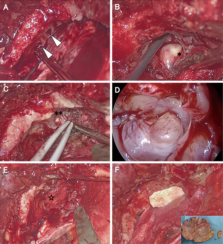
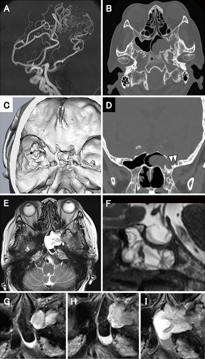
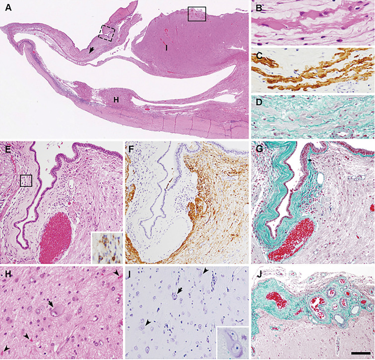
Meningoencephalocele in the lateral sphenoid sinus (SS) has been determined to be a rare entity often detected by cerebrospinal fluid (CSF) rhinorrhea. To date, the pathology of meningoencephalocele in the lateral SS has remained to be unclear in many cases. In this study, we report on a case of a 72-year-old woman with an arteriovenous malformation who presented with CSF rhinorrhea. Radiologic investigations revealed a left temporal meningoencephalocele in the lateral SS. We removed the meningoencephalocele and performed skull base repair, after which the CSF rhinorrhea resolved. Pathological examination showed congenital cortical abnormalities with dysmorphic neurons in various shapes and acquired chronic tissue alterations including fibrillary gliosis and scattered Rosenthal fibers. These findings may further aid in understanding the etiopathogenesis of meningoencephalocele in the lateral SS.


 分享
分享

 求助内容:
求助内容: 应助结果提醒方式:
 扫码关注我们
