{"title":"Endoscopic Removal of Pedunculated Leiomyoma of the Sigmoid Colon.","authors":"Harunobu Sato, Yoshihisa Mizuno, Tetsuya Tsukamoto, Tomoaki Ichikawa, Yoshihito Kotani, Katsuyuki Honda, Makoto Kuroda","doi":"10.1159/000368791","DOIUrl":null,"url":null,"abstract":"<p><strong>Background: </strong>The large bowel is a rare site for leiomyomas. Furthermore, a colonic pedunculated leiomyoma is very rare. Complete endoscopic removal of a colonic leiomyoma can be problematic because of its submucosal origin.</p><p><strong>Case report: </strong>We report a colonic pedunculated leiomyoma that was removed by endoscopic polypectomy without complications. A 74-year-old man was referred to our hospital because of constipation. Colonoscopy demonstrated a 1-cm pedunculated polyp that was connected to a minute stalk within the sigmoid colon. It was removed by snare polypectomy. Histopathological examination demonstrated normal mucosa overlying a well-circumscribed proliferation of eosinophilic spindle cells arising in association with the muscularis mucosae. Immunohistological findings were positive for desmin and smooth muscle actin. The polyp was diagnosed as a leiomyoma. More than 9 months later, the patient remains well, with no further symptoms.</p><p><strong>Conclusion: </strong>For small, pedunculated leiomyomas, endoscopic snare polypectomy is thought to be a useful approach for both treatment and diagnosis.</p>","PeriodicalId":49114,"journal":{"name":"Viszeralmedizin","volume":"30 6","pages":"427-9"},"PeriodicalIF":0.0000,"publicationDate":"2014-12-01","publicationTypes":"Journal Article","fieldsOfStudy":null,"isOpenAccess":false,"openAccessPdf":"https://sci-hub-pdf.com/10.1159/000368791","citationCount":"4","resultStr":null,"platform":"Semanticscholar","paperid":null,"PeriodicalName":"Viszeralmedizin","FirstCategoryId":"1085","ListUrlMain":"https://doi.org/10.1159/000368791","RegionNum":0,"RegionCategory":null,"ArticlePicture":[],"TitleCN":null,"AbstractTextCN":null,"PMCID":null,"EPubDate":"","PubModel":"","JCR":"","JCRName":"","Score":null,"Total":0}
引用次数: 4
Abstract
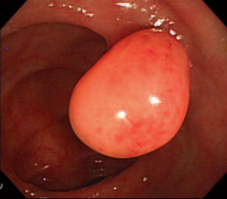
Background: The large bowel is a rare site for leiomyomas. Furthermore, a colonic pedunculated leiomyoma is very rare. Complete endoscopic removal of a colonic leiomyoma can be problematic because of its submucosal origin.
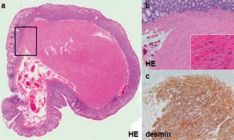
Case report: We report a colonic pedunculated leiomyoma that was removed by endoscopic polypectomy without complications. A 74-year-old man was referred to our hospital because of constipation. Colonoscopy demonstrated a 1-cm pedunculated polyp that was connected to a minute stalk within the sigmoid colon. It was removed by snare polypectomy. Histopathological examination demonstrated normal mucosa overlying a well-circumscribed proliferation of eosinophilic spindle cells arising in association with the muscularis mucosae. Immunohistological findings were positive for desmin and smooth muscle actin. The polyp was diagnosed as a leiomyoma. More than 9 months later, the patient remains well, with no further symptoms.
Conclusion: For small, pedunculated leiomyomas, endoscopic snare polypectomy is thought to be a useful approach for both treatment and diagnosis.


 分享
分享
 求助内容:
求助内容: 应助结果提醒方式:
 扫码关注我们
