Experimental Fusion of Contrast Enhanced High-Field Magnetic Resonance Imaging and High-Resolution Micro-Computed Tomography in Imaging the Mouse Inner Ear.
S Allen Counter, Peter Damberg, Sahar Nikkhou Aski, Kálmán Nagy, Cecilia Engmér Berglin, Göran Laurell
{"title":"Experimental Fusion of Contrast Enhanced High-Field Magnetic Resonance Imaging and High-Resolution Micro-Computed Tomography in Imaging the Mouse Inner Ear.","authors":"S Allen Counter, Peter Damberg, Sahar Nikkhou Aski, Kálmán Nagy, Cecilia Engmér Berglin, Göran Laurell","doi":"10.2174/1874440001509010007","DOIUrl":null,"url":null,"abstract":"<p><strong>Objective: </strong>Imaging cochlear, vestibular, and 8th cranial nerve abnormalities remains a challenge. In this study, the membranous and osseous labyrinths of the wild type mouse inner ear were examined using volumetric data from ultra high-field magnetic resonance imaging (MRI) with gadolinium contrast at 9.4 Tesla and high-resolution micro-computed tomography (µCT) to visualize the scalae and vestibular apparatus, and to establish imaging protocols and parameters for comparative analysis of the normal and mutant mouse inner ear.</p><p><strong>Methods: </strong>For in vivo MRI acquisition, animals were placed in a Milleped coil situated in the isocenter of a horizontal 9.4 T Varian magnet. For µCT examination, cone beam scans were performed ex vivo following MRI using the µCT component of a nanoScan PET/CT in vivo scanner.</p><p><strong>Results: </strong>The fusion of Gd enhanced high field MRI and high-resolution µCT scans revealed the dynamic membranous labyrinth of the perilymphatic fluid filled scala tympani and scala vestibule of the cochlea, and semicircular canals of the vestibular apparatus, within the µCT visualized contours of the contiguous osseous labyrinth. The ex vivo µCT segmentation revealed the surface contours and structural morphology of each cochlea turn and the semicircular canals in 3 planes.</p><p><strong>Conclusions: </strong>The fusion of ultra high-field MRI and high-resolution µCT imaging techniques were complementary, and provided high-resolution dynamic and static visualization of the complex morphological features of the normal mouse inner ear structures, which may offer a valuable approach for the investigation of cochlear and vestibular abnormalities that are associated with birth defects related to genetic inner ear disorders in humans.</p>","PeriodicalId":37431,"journal":{"name":"Open Neuroimaging Journal","volume":"9 ","pages":"7-12"},"PeriodicalIF":0.0000,"publicationDate":"2015-07-31","publicationTypes":"Journal Article","fieldsOfStudy":null,"isOpenAccess":false,"openAccessPdf":"https://ftp.ncbi.nlm.nih.gov/pub/pmc/oa_pdf/91/21/TONIJ-9-7.PMC4578136.pdf","citationCount":"9","resultStr":null,"platform":"Semanticscholar","paperid":null,"PeriodicalName":"Open Neuroimaging Journal","FirstCategoryId":"1085","ListUrlMain":"https://doi.org/10.2174/1874440001509010007","RegionNum":0,"RegionCategory":null,"ArticlePicture":[],"TitleCN":null,"AbstractTextCN":null,"PMCID":null,"EPubDate":"2015/1/1 0:00:00","PubModel":"eCollection","JCR":"Q4","JCRName":"Medicine","Score":null,"Total":0}
引用次数: 9
Abstract
Objective: Imaging cochlear, vestibular, and 8th cranial nerve abnormalities remains a challenge. In this study, the membranous and osseous labyrinths of the wild type mouse inner ear were examined using volumetric data from ultra high-field magnetic resonance imaging (MRI) with gadolinium contrast at 9.4 Tesla and high-resolution micro-computed tomography (µCT) to visualize the scalae and vestibular apparatus, and to establish imaging protocols and parameters for comparative analysis of the normal and mutant mouse inner ear.
Methods: For in vivo MRI acquisition, animals were placed in a Milleped coil situated in the isocenter of a horizontal 9.4 T Varian magnet. For µCT examination, cone beam scans were performed ex vivo following MRI using the µCT component of a nanoScan PET/CT in vivo scanner.
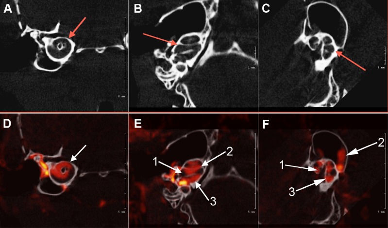
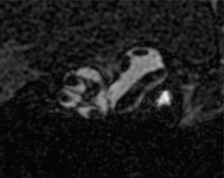
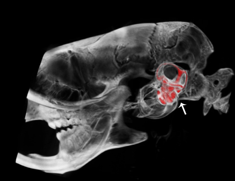
Results: The fusion of Gd enhanced high field MRI and high-resolution µCT scans revealed the dynamic membranous labyrinth of the perilymphatic fluid filled scala tympani and scala vestibule of the cochlea, and semicircular canals of the vestibular apparatus, within the µCT visualized contours of the contiguous osseous labyrinth. The ex vivo µCT segmentation revealed the surface contours and structural morphology of each cochlea turn and the semicircular canals in 3 planes.
Conclusions: The fusion of ultra high-field MRI and high-resolution µCT imaging techniques were complementary, and provided high-resolution dynamic and static visualization of the complex morphological features of the normal mouse inner ear structures, which may offer a valuable approach for the investigation of cochlear and vestibular abnormalities that are associated with birth defects related to genetic inner ear disorders in humans.
期刊介绍:
The Open Neuroimaging Journal is an Open Access online journal, which publishes research articles, reviews/mini-reviews, and letters in all important areas of brain function, structure and organization including neuroimaging, neuroradiology, analysis methods, functional MRI acquisition and physics, brain mapping, macroscopic level of brain organization, computational modeling and analysis, structure-function and brain-behavior relationships, anatomy and physiology, psychiatric diseases and disorders of the nervous system, use of imaging to the understanding of brain pathology and brain abnormalities, cognition and aging, social neuroscience, sensorimotor processing, communication and learning.


 分享
分享
 求助内容:
求助内容: 应助结果提醒方式:
 扫码关注我们
