Microvesicles derived from hypoxia/reoxygenation-treated human umbilical vein endothelial cells promote apoptosis and oxidative stress in H9c2 cardiomyocytes.
Qi Zhang, Man Shang, Mengxiao Zhang, Yao Wang, Yan Chen, Yanna Wu, Minglin Liu, Junqiu Song, Yanxia Liu
{"title":"Microvesicles derived from hypoxia/reoxygenation-treated human umbilical vein endothelial cells promote apoptosis and oxidative stress in H9c2 cardiomyocytes.","authors":"Qi Zhang, Man Shang, Mengxiao Zhang, Yao Wang, Yan Chen, Yanna Wu, Minglin Liu, Junqiu Song, Yanxia Liu","doi":"10.1186/s12860-016-0100-1","DOIUrl":null,"url":null,"abstract":"<p><strong>Background: </strong>Vascular endothelial dysfunction is the closely related determinant of ischemic heart disease (IHD). Endothelial dysfunction and ischemia/reperfusion injury (IRI) have been associated with an increase in microvesicles (MVs) in vivo. However, the potential contribution of endothelial microvesicles (EMVs) to myocardial damage is unclear. Here we aimed to investigate the role of EMVs derived from hypoxia/reoxygenation (H/R) -treated human umbilical vein endothelial cells (HUVECs) on cultured H9c2 cardiomyocytes.</p><p><strong>Results: </strong>H/R injury model was established to induce HUVECs to release H/R-EMVs. The H/R-EMVs from HUVECs were isolated from the conditioned culture medium and characterized. H9c2 cardiomyocytes were then incubated with 10, 30, 60 μg/mL H/R-EMVs for 6 h. We found that H9c2 cells treated by H/R-EMVs exhibited reduced cell viability, increased cell apoptosis and reactive oxygen species (ROS) production. Moreover mechanism studies demonstrated that H/R-EMVs could induce the phosphorylation of p38 and JNK1/2 in H9c2 cells in a dose-dependent manner. In addition, H/R-EMVs contained significantly higher level of ROS than EMVs generated from untreated HUVECs, which might be a direct source to trigger a cascade of myocardial damage.</p><p><strong>Conclusion: </strong>We showed that EMVs released during H/R injury are pro-apoptotic, pro-oxidative and directly pathogenic to cardiomyocytes in vitro. EMVs carry ROS and they may impair myocardium by promoting apoptosis and oxidative stress. These findings provide new insights into the pathogenesis of IRI.</p>","PeriodicalId":9051,"journal":{"name":"BMC Cell Biology","volume":"17 1","pages":"25"},"PeriodicalIF":0.0000,"publicationDate":"2016-06-23","publicationTypes":"Journal Article","fieldsOfStudy":null,"isOpenAccess":false,"openAccessPdf":"https://sci-hub-pdf.com/10.1186/s12860-016-0100-1","citationCount":"56","resultStr":null,"platform":"Semanticscholar","paperid":null,"PeriodicalName":"BMC Cell Biology","FirstCategoryId":"1085","ListUrlMain":"https://doi.org/10.1186/s12860-016-0100-1","RegionNum":0,"RegionCategory":null,"ArticlePicture":[],"TitleCN":null,"AbstractTextCN":null,"PMCID":null,"EPubDate":"","PubModel":"","JCR":"Q1","JCRName":"Biochemistry, Genetics and Molecular Biology","Score":null,"Total":0}
引用次数: 56
Abstract
Background: Vascular endothelial dysfunction is the closely related determinant of ischemic heart disease (IHD). Endothelial dysfunction and ischemia/reperfusion injury (IRI) have been associated with an increase in microvesicles (MVs) in vivo. However, the potential contribution of endothelial microvesicles (EMVs) to myocardial damage is unclear. Here we aimed to investigate the role of EMVs derived from hypoxia/reoxygenation (H/R) -treated human umbilical vein endothelial cells (HUVECs) on cultured H9c2 cardiomyocytes.
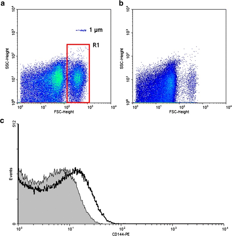
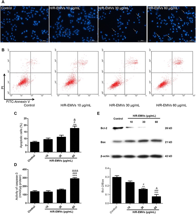
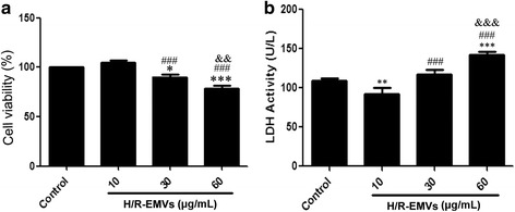
Results: H/R injury model was established to induce HUVECs to release H/R-EMVs. The H/R-EMVs from HUVECs were isolated from the conditioned culture medium and characterized. H9c2 cardiomyocytes were then incubated with 10, 30, 60 μg/mL H/R-EMVs for 6 h. We found that H9c2 cells treated by H/R-EMVs exhibited reduced cell viability, increased cell apoptosis and reactive oxygen species (ROS) production. Moreover mechanism studies demonstrated that H/R-EMVs could induce the phosphorylation of p38 and JNK1/2 in H9c2 cells in a dose-dependent manner. In addition, H/R-EMVs contained significantly higher level of ROS than EMVs generated from untreated HUVECs, which might be a direct source to trigger a cascade of myocardial damage.
Conclusion: We showed that EMVs released during H/R injury are pro-apoptotic, pro-oxidative and directly pathogenic to cardiomyocytes in vitro. EMVs carry ROS and they may impair myocardium by promoting apoptosis and oxidative stress. These findings provide new insights into the pathogenesis of IRI.
背景:血管内皮功能障碍与缺血性心脏病(IHD)密切相关。内皮功能障碍和缺血/再灌注损伤(IRI)与体内微囊泡(MVs)的增加有关。然而,内皮微泡(emv)对心肌损伤的潜在贡献尚不清楚。在这里,我们旨在研究缺氧/再氧化(H/R)处理的人脐静脉内皮细胞(HUVECs)衍生的emv对培养的H9c2心肌细胞的作用。结果:建立H/R损伤模型,诱导HUVECs释放H/R- emv。从条件培养基中分离HUVECs的H/ r - emv并对其进行鉴定。H9c2心肌细胞分别以10、30、60 μg/mL H/ r - emv孵育6小时,结果发现H/ r - emv处理的H9c2细胞活力降低,细胞凋亡增加,活性氧(ROS)产生增加。此外,机制研究表明,H/ r - emv可诱导H9c2细胞中p38和JNK1/2的磷酸化,并呈剂量依赖性。此外,H/ r - emv的ROS水平明显高于未经处理的huvec产生的emv,这可能是触发心肌损伤级联的直接来源。结论:H/R损伤释放的emv具有促凋亡、促氧化作用,对心肌细胞具有直接致病作用。emv携带ROS,可能通过促进细胞凋亡和氧化应激损害心肌。这些发现为IRI的发病机制提供了新的见解。
期刊介绍:
BMC Molecular and Cell Biology, formerly known as BMC Cell Biology, is an open access journal that considers articles on all aspects of both eukaryotic and prokaryotic cell and molecular biology, including structural and functional cell biology, DNA and RNA in a cellular context and biochemistry, as well as research using both the experimental and theoretical aspects of physics to study biological processes and investigations into the structure of biological macromolecules.


 分享
分享
 求助内容:
求助内容: 应助结果提醒方式:
 扫码关注我们
