R Witt, A Weigand, A M Boos, A Cai, D Dippold, A R Boccaccini, D W Schubert, M Hardt, C Lange, A Arkudas, R E Horch, J P Beier
{"title":"Mesenchymal stem cells and myoblast differentiation under HGF and IGF-1 stimulation for 3D skeletal muscle tissue engineering.","authors":"R Witt, A Weigand, A M Boos, A Cai, D Dippold, A R Boccaccini, D W Schubert, M Hardt, C Lange, A Arkudas, R E Horch, J P Beier","doi":"10.1186/s12860-017-0131-2","DOIUrl":null,"url":null,"abstract":"<p><strong>Background: </strong>Volumetric muscle loss caused by trauma or after tumour surgery exceeds the natural regeneration capacity of skeletal muscle. Hence, the future goal of tissue engineering (TE) is the replacement and repair of lost muscle tissue by newly generating skeletal muscle combining different cell sources, such as myoblasts and mesenchymal stem cells (MSCs), within a three-dimensional matrix. Latest research showed that seeding skeletal muscle cells on aligned constructs enhance the formation of myotubes as well as cell alignment and may provide a further step towards the clinical application of engineered skeletal muscle. In this study the myogenic differentiation potential of MSCs upon co-cultivation with myoblasts and under stimulation with hepatocyte growth factor (HGF) and insulin-like growth factor-1 (IGF-1) was evaluated. We further analysed the behaviour of MSC-myoblast co-cultures in different 3D matrices.</p><p><strong>Results: </strong>Primary rat myoblasts and rat MSCs were mono- and co-cultivated for 2, 7 or 14 days. The effect of different concentrations of HGF and IGF-1 alone, as well as in combination, on myogenic differentiation was analysed using microscopy, multicolour flow cytometry and real-time PCR. Furthermore, the influence of different three-dimensional culture models, such as fibrin, fibrin-collagen-I gels and parallel aligned electrospun poly-ε-caprolacton collagen-I nanofibers, on myogenic differentiation was analysed. MSCs could be successfully differentiated into the myogenic lineage both in mono- and in co-cultures independent of HGF and IGF-1 stimulation by expressing desmin, myocyte enhancer factor 2, myosin heavy chain 2 and alpha-sarcomeric actinin. An increased expression of different myogenic key markers could be observed under HGF and IGF-1 stimulation. Even though, stimulation with HGF/IGF-1 does not seem essential for sufficient myogenic differentiation. Three-dimensional cultivation in fibrin-collagen-I gels induced higher levels of myogenic differentiation compared with two-dimensional experiments. Cultivation on poly-ε-caprolacton-collagen-I nanofibers induced parallel alignment of cells and positive expression of desmin.</p><p><strong>Conclusions: </strong>In this study, we were able to myogenically differentiate MSC upon mono- and co-cultivation with myoblasts. The addition of HGF/IGF-1 might not be essential for achieving successful myogenic differentiation. Furthermore, with the development of a biocompatible nanofiber scaffold we established the basis for further experiments aiming at the generation of functional muscle tissue.</p>","PeriodicalId":9051,"journal":{"name":"BMC Cell Biology","volume":"18 1","pages":"15"},"PeriodicalIF":0.0000,"publicationDate":"2017-02-28","publicationTypes":"Journal Article","fieldsOfStudy":null,"isOpenAccess":false,"openAccessPdf":"https://sci-hub-pdf.com/10.1186/s12860-017-0131-2","citationCount":"70","resultStr":null,"platform":"Semanticscholar","paperid":null,"PeriodicalName":"BMC Cell Biology","FirstCategoryId":"1085","ListUrlMain":"https://doi.org/10.1186/s12860-017-0131-2","RegionNum":0,"RegionCategory":null,"ArticlePicture":[],"TitleCN":null,"AbstractTextCN":null,"PMCID":null,"EPubDate":"","PubModel":"","JCR":"Q1","JCRName":"Biochemistry, Genetics and Molecular Biology","Score":null,"Total":0}
引用次数: 70
Abstract
Background: Volumetric muscle loss caused by trauma or after tumour surgery exceeds the natural regeneration capacity of skeletal muscle. Hence, the future goal of tissue engineering (TE) is the replacement and repair of lost muscle tissue by newly generating skeletal muscle combining different cell sources, such as myoblasts and mesenchymal stem cells (MSCs), within a three-dimensional matrix. Latest research showed that seeding skeletal muscle cells on aligned constructs enhance the formation of myotubes as well as cell alignment and may provide a further step towards the clinical application of engineered skeletal muscle. In this study the myogenic differentiation potential of MSCs upon co-cultivation with myoblasts and under stimulation with hepatocyte growth factor (HGF) and insulin-like growth factor-1 (IGF-1) was evaluated. We further analysed the behaviour of MSC-myoblast co-cultures in different 3D matrices.
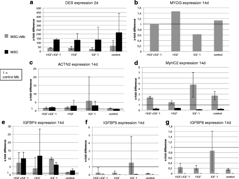
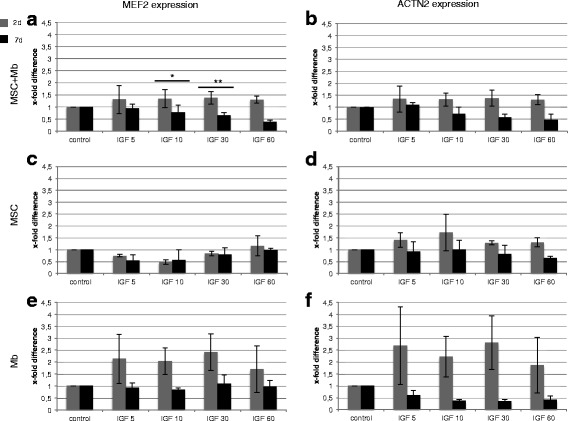
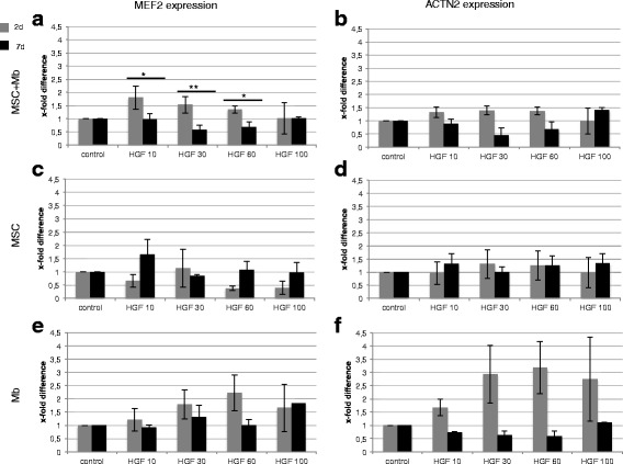
Results: Primary rat myoblasts and rat MSCs were mono- and co-cultivated for 2, 7 or 14 days. The effect of different concentrations of HGF and IGF-1 alone, as well as in combination, on myogenic differentiation was analysed using microscopy, multicolour flow cytometry and real-time PCR. Furthermore, the influence of different three-dimensional culture models, such as fibrin, fibrin-collagen-I gels and parallel aligned electrospun poly-ε-caprolacton collagen-I nanofibers, on myogenic differentiation was analysed. MSCs could be successfully differentiated into the myogenic lineage both in mono- and in co-cultures independent of HGF and IGF-1 stimulation by expressing desmin, myocyte enhancer factor 2, myosin heavy chain 2 and alpha-sarcomeric actinin. An increased expression of different myogenic key markers could be observed under HGF and IGF-1 stimulation. Even though, stimulation with HGF/IGF-1 does not seem essential for sufficient myogenic differentiation. Three-dimensional cultivation in fibrin-collagen-I gels induced higher levels of myogenic differentiation compared with two-dimensional experiments. Cultivation on poly-ε-caprolacton-collagen-I nanofibers induced parallel alignment of cells and positive expression of desmin.
Conclusions: In this study, we were able to myogenically differentiate MSC upon mono- and co-cultivation with myoblasts. The addition of HGF/IGF-1 might not be essential for achieving successful myogenic differentiation. Furthermore, with the development of a biocompatible nanofiber scaffold we established the basis for further experiments aiming at the generation of functional muscle tissue.
期刊介绍:
BMC Molecular and Cell Biology, formerly known as BMC Cell Biology, is an open access journal that considers articles on all aspects of both eukaryotic and prokaryotic cell and molecular biology, including structural and functional cell biology, DNA and RNA in a cellular context and biochemistry, as well as research using both the experimental and theoretical aspects of physics to study biological processes and investigations into the structure of biological macromolecules.


 分享
分享
 求助内容:
求助内容: 应助结果提醒方式:
 扫码关注我们
